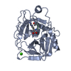
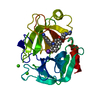
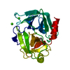
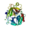
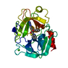
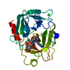
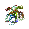
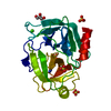
SHEET DETERMINATION METHOD: DSSP THE SHEETS PRESENTED AS "AB" IN EACH CHAIN ON SHEET RECORDS BELOW ... SHEET DETERMINATION METHOD: DSSP THE SHEETS PRESENTED AS "AB" IN EACH CHAIN ON SHEET RECORDS BELOW IS ACTUALLY AN 6-STRANDED BARREL THIS IS REPRESENTED BY A 7-STRANDED SHEET IN WHICH THE FIRST AND LAST STRANDS ARE IDENTICAL.
Mass: 18.015 Da / Num. of mol.: 110 / Source method: isolated from a natural source / Formula: H2O
-
Details
Has protein modification
Y
Sequence details
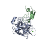
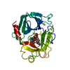
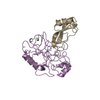
VERONICA HEDERIFOLIA TRYPSIN INHIBITOR SEQUENCE IS NOT PRESENT IN ANY SEQUENCE DATABASE. WE WILL ...VERONICA HEDERIFOLIA TRYPSIN INHIBITOR SEQUENCE IS NOT PRESENT IN ANY SEQUENCE DATABASE. WE WILL SUBMIT THE SEQUENCE ON PUBLICATION.
-
Experimental details
-
Experiment
Experiment
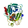
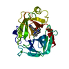
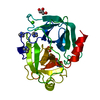
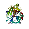
Method: X-RAY DIFFRACTION / Number of used crystals: 1
-
Sample preparation
Crystal
Density Matthews: 2.58 Å3/Da / Density % sol: 51.96 % / Description: NONE
Crystal grow
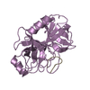
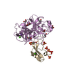
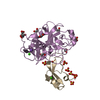
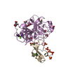
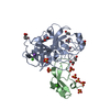
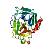
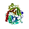
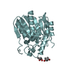
pH: 8 Details: CRYSTALS OF TRYPSIN WERE GROWN IN 2.5M AMMONIUM SULPHATE, 6MM CALCIUM CHLORIDE, 0.1M TRIS PH 8.15, 60MM BENZAMIDINE. THEY WERE BACKSOAKED IN 0.1M NA PHOSPHATE PH 5.8, 2.5M AMMONIUM SULPHATE ...Details: CRYSTALS OF TRYPSIN WERE GROWN IN 2.5M AMMONIUM SULPHATE, 6MM CALCIUM CHLORIDE, 0.1M TRIS PH 8.15, 60MM BENZAMIDINE. THEY WERE BACKSOAKED IN 0.1M NA PHOSPHATE PH 5.8, 2.5M AMMONIUM SULPHATE TO REMOVE BENZAMIDINE. CRYSTALS WERE MOVED TO 2.5M AMMONIUM SULPHATE, 1MM CALCIUM CHLORDIE, 0.1M TRIS PH 8 AND PEPTIDE INHIBITOR ADDED AT 10MM AND INCUBATED FOR 16 HOURS.
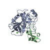
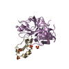
Resolution: 2.25→47.73 Å / Cor.coef. Fo:Fc: 0.953 / Cor.coef. Fo:Fc free: 0.917 / SU B: 12.837 / SU ML: 0.176 / TLS residual ADP flag: LIKELY RESIDUAL / Cross valid method: THROUGHOUT / ESU R: 0.278 / ESU R Free: 0.228 / Stereochemistry target values: MAXIMUM LIKELIHOOD Details: HYDROGENS HAVE BEEN ADDED IN THE RIDING POSITIONS. RESIDUES 1-6, 16-17 AND 30-34 OF THE VERONICA HEDERIFOLIA INHIBITOR (CHAIN B) ARE DISORDERED AND NOT INCLUDED IN THE MODEL.
Rfactor
Num. reflection
% reflection
Selection details
Rfree
0.253
661
5 %
RANDOM
Rwork
0.194
-
-
-
obs
0.196
12681
96.9 %
-
Solvent computation
Ion probe radii: 0.8 Å / Shrinkage radii: 0.8 Å / VDW probe radii: 1.4 Å / Solvent model: BABINET MODEL WITH MASK
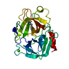
 Movie
Movie Controller

 Yorodumi
 Open data
 Basic information
 Components
 Keywords
 Function and homology information
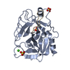
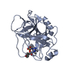
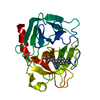
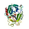
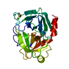
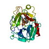
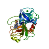
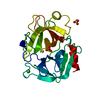
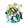
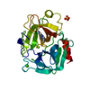
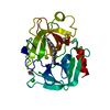
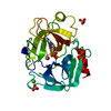
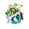
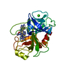
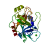
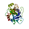
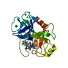
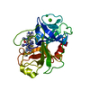
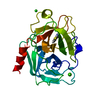
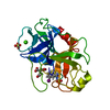
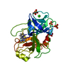
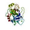
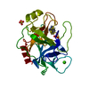
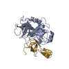
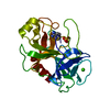
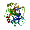
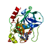
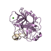
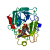
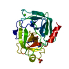
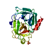
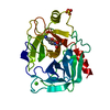
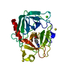
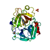
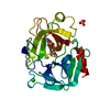
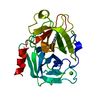
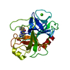
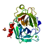
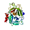
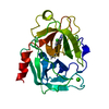
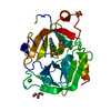
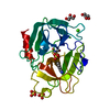
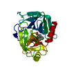
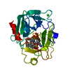
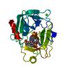
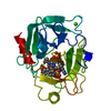
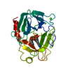
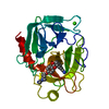
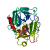
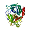
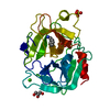
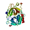
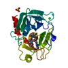
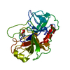
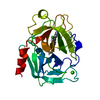
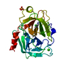
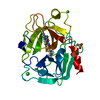
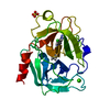
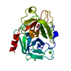
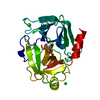
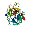
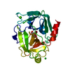
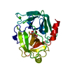
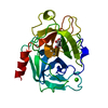
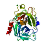
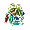
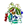
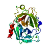
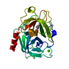
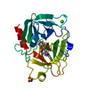
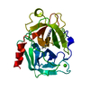
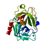
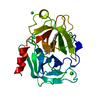
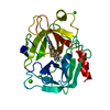
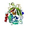
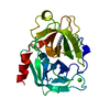
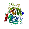
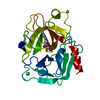
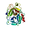
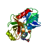
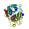
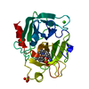
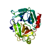
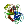
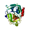
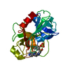
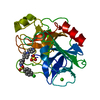
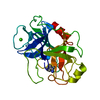
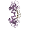
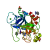
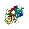
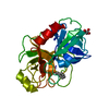
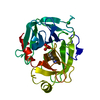
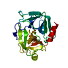
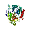
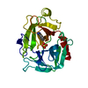
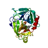
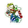
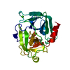
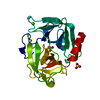
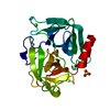
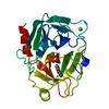
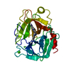
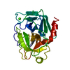
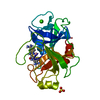
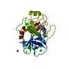
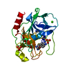
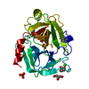
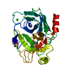
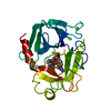
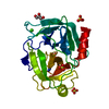
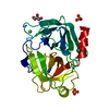
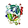
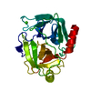
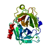
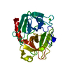
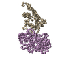
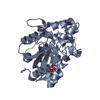
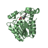
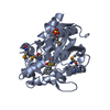
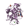
 VERONICA HEDERIFOLIA (ivy-leaved speedwell)
 X-RAY DIFFRACTION /
  Authors
 Citation
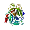
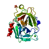
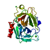
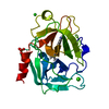
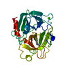
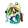
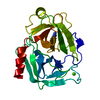
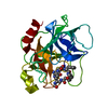
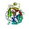
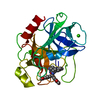
 Structure visualization
 Downloads & links
 Other downloads


 PDBj
PDBj

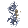
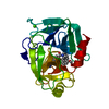
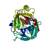
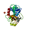
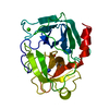
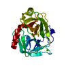
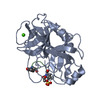
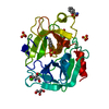
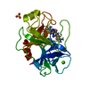
 Assembly
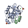


 Mass: 96.063 Da / Num. of mol.: 2 / Source method: obtained synthetically / Formula: SO4
 Mass: 40.078 Da / Num. of mol.: 1 / Source method: obtained synthetically / Formula: Ca
 Mass: 92.094 Da / Num. of mol.: 1 / Source method: obtained synthetically / Formula: C3H8O3
 Sample preparation
 / Beamline: PX14.1 / Wavelength: 1.488
  Processing