 Movie
Movie Controller

[English] 日本語
 Yorodumi
- PDB-1ejm: CRYSTAL STRUCTURE OF THE BPTI ALA16LEU MUTANT IN COMPLEX WITH BOV... -
+ Open data

- Basic information
| Entry | Database: PDB / ID: 1ejm | ||||||
|---|---|---|---|---|---|---|---|
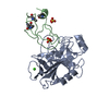
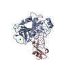
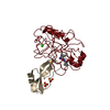
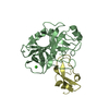
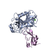
| Title | CRYSTAL STRUCTURE OF THE BPTI ALA16LEU MUTANT IN COMPLEX WITH BOVINE TRYPSIN | ||||||
 Components Components |
| ||||||
 Keywords Keywords | HYDROLASE/INHIBITOR / Complex / HYDROLASE-INHIBITOR COMPLEX | ||||||
| Function / homology |  Function and homology information Function and homology informationsulfate binding / negative regulation of platelet aggregation / zymogen binding / potassium channel inhibitor activity / molecular function inhibitor activity / negative regulation of thrombin-activated receptor signaling pathway / trypsin / serpin family protein binding / serine protease inhibitor complex / digestion ...sulfate binding / negative regulation of platelet aggregation / zymogen binding / potassium channel inhibitor activity / molecular function inhibitor activity / negative regulation of thrombin-activated receptor signaling pathway / trypsin / serpin family protein binding / serine protease inhibitor complex / digestion / serine-type endopeptidase inhibitor activity / protease binding / endopeptidase activity / serine-type endopeptidase activity / calcium ion binding / proteolysis / : / metal ion binding Similarity search - Function | ||||||
| Biological species |  | ||||||
| Method |  X-RAY DIFFRACTION / SYNCHROTRON / Resolution: 1.85 Å X-RAY DIFFRACTION / SYNCHROTRON / Resolution: 1.85 Å | ||||||
 Authors Authors | Otlewski, J. / Smalas, A. / Helland, R. / Grzesiak, A. / Krowarsch, D. | ||||||
 Citation Citation | Journal: J.Mol.Biol. / Year: 2000 Title: Substitutions at the P(1) position in BPTI strongly affect the association energy with serine proteinases. Authors: Grzesiak, A. / Helland, R. / Smalas, A.O. / Krowarsch, D. / Dadlez, M. / Otlewski, J. | ||||||
| History |
|
- Structure visualization
| Structure viewer | Molecule: MolmilJmol/JSmol |
|---|
- Downloads & links
-Download
| PDBx/mmCIF format | 1ejm.cif.gz | 181 KB | Display | PDBx/mmCIF format |
|---|---|---|---|---|
| PDB format | pdb1ejm.ent.gz | 143.4 KB | Display | PDB format |
| PDBx/mmJSON format | 1ejm.json.gz | Tree view | PDBx/mmJSON format | |
| Others |  Other downloads Other downloads |
-Validation report
| Arichive directory | https://data.pdbj.org/pub/pdb/validation_reports/ej/1ejmftp://data.pdbj.org/pub/pdb/validation_reports/ej/1ejm | HTTPS FTP |
|---|
-Related structure data
| Similar structure data |
|---|

-Links
 PDBj
PDBj


- Assembly
| Deposited unit | 
| ||||||||
|---|---|---|---|---|---|---|---|---|---|
| 1 | 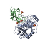
| ||||||||
| 2 | 
| ||||||||
| 3 | 
| ||||||||
| Unit cell |
| ||||||||
| Details | There are 3 complex molecules in the asymmetric unit. |
-Components
| #1: Protein | Mass: 23324.287 Da / Num. of mol.: 3 / Source method: isolated from a natural source / Details: PURCHASED FROM SIGMA / Source: (natural) #2: Protein | Mass: 6579.624 Da / Num. of mol.: 3 / Mutation: K15R, A16L, M52L Source method: isolated from a genetically manipulated source Source: (gene. exp.)  #3: Chemical | ChemComp-SO4 /   Mass: 96.063 Da / Num. of mol.: 12 / Source method: obtained synthetically / Formula: SO4 Mass: 96.063 Da / Num. of mol.: 12 / Source method: obtained synthetically / Formula: SO4#4: Water | ChemComp-HOH / |  Mass: 18.015 Da / Num. of mol.: 611 / Source method: isolated from a natural source / Formula: H2O Mass: 18.015 Da / Num. of mol.: 611 / Source method: isolated from a natural source / Formula: H2OHas protein modification | Y | |
|---|
-Experimental details
-Experiment
| Experiment | Method: X-RAY DIFFRACTION / Number of used crystals: 1 |
|---|
- Sample preparation
| Crystal | Density Matthews: 3.88 Å3/Da / Density % sol: 68.34 % | ||||||||||||||||||||
|---|---|---|---|---|---|---|---|---|---|---|---|---|---|---|---|---|---|---|---|---|---|
| Crystal grow | Temperature: 310 K / Method: vapor diffusion, hanging drop / pH: 7.5 Details: 48% saturated ammonium sulphate, 0.1 M Hepes buffer, pH 7.5, VAPOR DIFFUSION, HANGING DROP, temperature 37K | ||||||||||||||||||||
| Crystal grow | *PLUS Details: Helland, R., (1999) J. Mol. Biol., 287, 923. | ||||||||||||||||||||
| Components of the solutions | *PLUS
|
-Data collection
| Diffraction | Mean temperature: 120 K |
|---|---|
| Diffraction source | Source: SYNCHROTRON / Site: ESRF  / Type: ESRF / Wavelength: 0.93 / Type: ESRF / Wavelength: 0.93 |
| Detector | Type: ADSC QUANTUM 4 / Detector: CCD / Date: Jun 18, 1999 |
| Radiation | Protocol: SINGLE WAVELENGTH / Monochromatic (M) / Laue (L): M / Scattering type: x-ray |
| Radiation wavelength | Wavelength: 0.93 Å / Relative weight: 1 |
| Reflection | Resolution: 1.85→15 Å / Num. all: 1193911 / Num. obs: 119391 / % possible obs: 99.4 % / Observed criterion σ(F): 0 / Observed criterion σ(I): 0 / Redundancy: 7.4 % / Biso Wilson estimate: 30.3 Å2 / Rmerge(I) obs: 0.071 / Net I/σ(I): 5.4 |
| Reflection shell | Resolution: 1.85→1.95 Å / Redundancy: 1.8 % / Rmerge(I) obs: 0.412 / % possible all: 99.3 |
| Reflection | *PLUS Num. measured all: 1360301 |
- Processing
| Software |
| ||||||||||||||||||||
|---|---|---|---|---|---|---|---|---|---|---|---|---|---|---|---|---|---|---|---|---|---|
| Refinement | Resolution: 1.85→15 Å / σ(F): 0 / σ(I): 0 / Stereochemistry target values: Eng & Huber
| ||||||||||||||||||||
| Refinement step | Cycle: LAST / Resolution: 1.85→15 Å
| ||||||||||||||||||||
| Refine LS restraints |
| ||||||||||||||||||||
| Software | *PLUS Name: CNS / Classification: refinement | ||||||||||||||||||||
| Refinement | *PLUS Lowest resolution: 15 Å / σ(F): 0 / Rfactor obs: 0.211 | ||||||||||||||||||||
| Solvent computation | *PLUS | ||||||||||||||||||||
| Displacement parameters | *PLUS Biso mean: 30.3 Å2 | ||||||||||||||||||||
| Refine LS restraints | *PLUS
|
