 Movie
Movie Controller

[English] 日本語
 Yorodumi
- PDB-1tfx: COMPLEX OF THE SECOND KUNITZ DOMAIN OF TISSUE FACTOR PATHWAY INHI... -
+ Open data

- Basic information
| Entry | Database: PDB / ID: 1tfx | ||||||
|---|---|---|---|---|---|---|---|
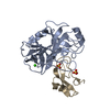
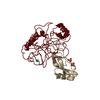
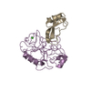
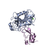
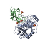
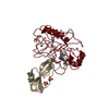
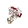
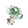
| Title | COMPLEX OF THE SECOND KUNITZ DOMAIN OF TISSUE FACTOR PATHWAY INHIBITOR WITH PORCINE TRYPSIN | ||||||
 Components Components |
| ||||||
 Keywords Keywords | COMPLEX (SERINE PROTEASE/INHIBITOR) / COMPLEX (SERINE PROTEASE-INHIBITOR) / HYDROLASE / INHIBITOR / BLOOD COAGULATION / COMPLEX (SERINE PROTEASE-INHIBITOR) complex | ||||||
| Function / homology |  Function and homology information Function and homology information: / cellular response to steroid hormone stimulus / endopeptidase inhibitor activity / trypsin / negative regulation of blood coagulation / digestion / side of membrane / serine-type endopeptidase inhibitor activity / caveola / blood coagulation ...: / cellular response to steroid hormone stimulus / endopeptidase inhibitor activity / trypsin / negative regulation of blood coagulation / digestion / side of membrane / serine-type endopeptidase inhibitor activity / caveola / blood coagulation / serine-type endopeptidase activity / cell surface / proteolysis / : / extracellular region / metal ion binding / plasma membrane Similarity search - Function | ||||||
| Biological species |   Homo sapiens (human) Homo sapiens (human) | ||||||
| Method |  X-RAY DIFFRACTION / MOLECULAR REPLACEMENT / Resolution: 2.6 Å X-RAY DIFFRACTION / MOLECULAR REPLACEMENT / Resolution: 2.6 Å | ||||||
 Authors Authors | Stubbs, M.T. / Huber, R. | ||||||
 Citation Citation | Journal: J.Mol.Biol. / Year: 1997 Title: The second Kunitz domain of human tissue factor pathway inhibitor: cloning, structure determination and interaction with factor Xa. Authors: Burgering, M.J. / Orbons, L.P. / van der Doelen, A. / Mulders, J. / Theunissen, H.J. / Grootenhuis, P.D. / Bode, W. / Huber, R. / Stubbs, M.T. #1: Journal: J.Biol.Chem. / Year: 1997Title: The Three-Dimensional Structure of Recombinant Leech-Derived Tryptase Inhibitor in Complex with Trypsin. Implications for the Structure of Human Mast Cell Tryptase and its Inhibition Authors: Stubbs, M.T. / Morenweiser, R. / Sturzebecher, J. / Bauer, M. / Bode, W. / Huber, R. / Piechottka, G.P. / Matschiner, G. / Sommerhoff, C.P. / Fritz, H. / Auerswald, E.A. #2: Journal: J.Biol.Chem. / Year: 1996Title: X-Ray Structure of Active Site-Inhibited Clotting Factor Xa. Implications for Drug Design and Substrate Recognition Authors: Brandstetter, H. / Kuhne, A. / Bode, W. / Huber, R. / Von Der Saal, W. / Wirthensohn, K. / Engh, R.A. #3: Journal: Curr.Pharm.Des. / Year: 1996Title: Structural Aspects of Factor Xa Inhibition Authors: Stubbs II, M.T. #4: Journal: Embo J. / Year: 1996Title: The Ornithodorin-Thrombin Crystal Structure, a Key to the Tap Enigma? Authors: Van De Locht, A. / Stubbs, M.T. / Bode, W. / Friedrich, T. / Bollschweiler, C. / Hoffken, W. / Huber, R. #5: Journal: FEBS Lett. / Year: 1995Title: Crystal Structures of Factor Xa Specific Inhibitors in Complex with Trypsin: Structural Grounds for Inhibition of Factor Xa and Selectivity Against Thrombin Authors: Stubbs, M.T. / Huber, R. / Bode, W. #6: Journal: J.Mol.Biol. / Year: 1993Title: Structure of Human Des(1-45) Factor Xa at 2.2 A Resolution Authors: Padmanabhan, K. / Padmanabhan, K.P. / Tulinsky, A. / Park, C.H. / Bode, W. / Huber, R. / Blankenship, D.T. / Cardin, A.D. / Kisiel, W. | ||||||
| History |
|
- Structure visualization
| Structure viewer | Molecule: MolmilJmol/JSmol |
|---|
- Downloads & links
-Download
| PDBx/mmCIF format | 1tfx.cif.gz | 119.5 KB | Display | PDBx/mmCIF format |
|---|---|---|---|---|
| PDB format | pdb1tfx.ent.gz | 92.4 KB | Display | PDB format |
| PDBx/mmJSON format | 1tfx.json.gz | Tree view | PDBx/mmJSON format | |
| Others |  Other downloads Other downloads |
-Validation report
| Arichive directory | https://data.pdbj.org/pub/pdb/validation_reports/tf/1tfxftp://data.pdbj.org/pub/pdb/validation_reports/tf/1tfx | HTTPS FTP |
|---|
-Related structure data
| Related structure data |  1adzC  1ldtS S: Starting model for refinement C: citing same article ( |
|---|---|
| Similar structure data |


-Links
 PDBj
PDBj


- Assembly
| Deposited unit | 
| ||||||||
|---|---|---|---|---|---|---|---|---|---|
| 1 | 
| ||||||||
| 2 | 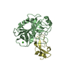
| ||||||||
| Unit cell |
| ||||||||
| Noncrystallographic symmetry (NCS) | NCS oper: (Code: given Matrix: (0.9999, -0.0114, 0.0008), Vector: |
-Components
| #1: Protein | Mass: 23493.496 Da / Num. of mol.: 2 Source method: isolated from a genetically manipulated source Source: (gene. exp.)  #2: Protein | Mass: 6853.665 Da / Num. of mol.: 2 / Fragment: FACTOR XA-BINDING DOMAIN, DOMAIN II Source method: isolated from a genetically manipulated source Source: (gene. exp.) Homo sapiens (human) / Tissue: BLOOD / Organ: BLOOD / Plasmid: PFLAG / Production host: #3: Chemical |   Mass: 40.078 Da / Num. of mol.: 2 / Source method: obtained synthetically / Formula: Ca Mass: 40.078 Da / Num. of mol.: 2 / Source method: obtained synthetically / Formula: Ca#4: Water | ChemComp-HOH / |  Mass: 18.015 Da / Num. of mol.: 143 / Source method: isolated from a natural source / Formula: H2O Mass: 18.015 Da / Num. of mol.: 143 / Source method: isolated from a natural source / Formula: H2OCompound details | THIS STRUCTURE IS PART OF A MULTIDISCIPLINARY STUDY INTO THE STRUCTURE AND FUNCTION OF THE SECOND ...THIS STRUCTURE IS PART OF A MULTIDISCI | Has protein modification | Y | |
|---|
-Experimental details
-Experiment
| Experiment | Method: X-RAY DIFFRACTION / Number of used crystals: 1 |
|---|
- Sample preparation
| Crystal | Density Matthews: 2.29 Å3/Da / Density % sol: 46 % |
|---|---|
| Crystal grow | pH: 8 / Details: pH 8.0 |
| Crystal grow | *PLUS Method: vapor diffusion, hanging drop |
| Components of the solutions | *PLUS Conc.: 1 M / Common name: Na/K phosphate |
-Data collection
| Diffraction | Mean temperature: 287 K |
|---|---|
| Diffraction source | Source: ROTATING ANODE / Type: RIGAKU / Wavelength: 1.5418 |
| Detector | Type: SIEMENS / Detector: AREA DETECTOR / Date: Jan 1, 1995 / Details: MIRRORS |
| Radiation | Monochromator: NI FILTER / Monochromatic (M) / Laue (L): M / Scattering type: x-ray |
| Radiation wavelength | Wavelength: 1.5418 Å / Relative weight: 1 |
| Reflection | Highest resolution: 2.6 Å / Num. obs: 17364 / % possible obs: 97 % / Observed criterion σ(I): 2 / Redundancy: 1.9 % / Rmerge(I) obs: 0.102 / Rsym value: 0.102 / Net I/σ(I): 9 |
| Reflection shell | Resolution: 2.6→2.7 Å / Redundancy: 1.5 % / Rmerge(I) obs: 0.3 / Mean I/σ(I) obs: 2 / Rsym value: 0.3 / % possible all: 97.6 |
| Reflection | *PLUS Lowest resolution: 9999 Å / Num. measured all: 32380 |
| Reflection shell | *PLUS % possible obs: 97.6 % |
- Processing
| Software |
| ||||||||||||||||||||||||||||||||||||||||||||||||||||||||||||
|---|---|---|---|---|---|---|---|---|---|---|---|---|---|---|---|---|---|---|---|---|---|---|---|---|---|---|---|---|---|---|---|---|---|---|---|---|---|---|---|---|---|---|---|---|---|---|---|---|---|---|---|---|---|---|---|---|---|---|---|---|---|
| Refinement | Method to determine structure: MOLECULAR REPLACEMENT Starting model: PORCINE TRYPSIN MODEL FROM LDTI TRYPSIN (PDB ENTRY 1LDT) Resolution: 2.6→6 Å / Data cutoff high absF: 10000000 / Data cutoff low absF: 0.001 / σ(F): 0
| ||||||||||||||||||||||||||||||||||||||||||||||||||||||||||||
| Refine analyze | Luzzati d res low obs: 6 Å | ||||||||||||||||||||||||||||||||||||||||||||||||||||||||||||
| Refinement step | Cycle: LAST / Resolution: 2.6→6 Å
| ||||||||||||||||||||||||||||||||||||||||||||||||||||||||||||
| Refine LS restraints |
| ||||||||||||||||||||||||||||||||||||||||||||||||||||||||||||
| LS refinement shell | Resolution: 2.6→2.64 Å / Total num. of bins used: 20
| ||||||||||||||||||||||||||||||||||||||||||||||||||||||||||||
| Xplor file |
| ||||||||||||||||||||||||||||||||||||||||||||||||||||||||||||
| Software | *PLUS Name: X-PLOR / Version: 3.1 / Classification: refinement | ||||||||||||||||||||||||||||||||||||||||||||||||||||||||||||
| Refinement | *PLUS | ||||||||||||||||||||||||||||||||||||||||||||||||||||||||||||
| Solvent computation | *PLUS | ||||||||||||||||||||||||||||||||||||||||||||||||||||||||||||
| Displacement parameters | *PLUS | ||||||||||||||||||||||||||||||||||||||||||||||||||||||||||||
| Refine LS restraints | *PLUS
| ||||||||||||||||||||||||||||||||||||||||||||||||||||||||||||
| LS refinement shell | *PLUS Rfactor obs: 0.212 |
