 Movie
Movie Controller

[English] 日本語
 Yorodumi
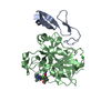
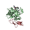
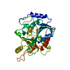
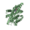
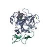
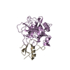
- PDB-4wxv: Human cationic trypsin K97D mutant in complex with bovine pancrea... -
+ Open data

- Basic information
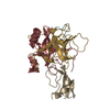
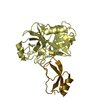
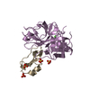
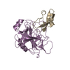
| Entry | Database: PDB / ID: 4wxv | ||||||
|---|---|---|---|---|---|---|---|
| Title | Human cationic trypsin K97D mutant in complex with bovine pancreatic trypsin inhibitor (BPTI) | ||||||
 Components Components |
| ||||||
 Keywords Keywords | Hydrolase/Hydrolase Inhibitor / trypsin inhibitor / BPTI / Hydrolase-Hydrolase Inhibitor complex | ||||||
| Function / homology |  Function and homology information Function and homology informationUptake of dietary cobalamins into enterocytes / Developmental Lineage of Pancreatic Acinar Cells / sulfate binding / negative regulation of platelet aggregation / zymogen binding / potassium channel inhibitor activity / molecular function inhibitor activity / Activation of Matrix Metalloproteinases / negative regulation of thrombin-activated receptor signaling pathway / extracellular matrix disassembly ...Uptake of dietary cobalamins into enterocytes / Developmental Lineage of Pancreatic Acinar Cells / sulfate binding / negative regulation of platelet aggregation / zymogen binding / potassium channel inhibitor activity / molecular function inhibitor activity / Activation of Matrix Metalloproteinases / negative regulation of thrombin-activated receptor signaling pathway / extracellular matrix disassembly / trypsin / serine protease inhibitor complex / digestion / serine-type endopeptidase inhibitor activity / extracellular matrix / protease binding / blood microparticle / serine-type endopeptidase activity / calcium ion binding / proteolysis / : / extracellular region / metal ion binding Similarity search - Function | ||||||
| Biological species |  Homo sapiens (human) Homo sapiens (human) | ||||||
| Method |  X-RAY DIFFRACTION / SYNCHROTRON / MOLECULAR REPLACEMENT / Resolution: 2.1 Å X-RAY DIFFRACTION / SYNCHROTRON / MOLECULAR REPLACEMENT / Resolution: 2.1 Å | ||||||
 Authors Authors | Alloy, A. / Kayode, O. / Soares, A.S. / Wang, R. / Radisky, E.S. | ||||||
| Funding support |  United States, 1items United States, 1items
| ||||||
 Citation Citation | Journal: J.Biol.Chem. / Year: 2015 Title: Mesotrypsin Has Evolved Four Unique Residues to Cleave Trypsin Inhibitors as Substrates. Authors: Alloy, A.P. / Kayode, O. / Wang, R. / Hockla, A. / Soares, A.S. / Radisky, E.S. | ||||||
| History |
|
- Structure visualization
| Structure viewer | Molecule: MolmilJmol/JSmol |
|---|
- Downloads & links
-Download
| PDBx/mmCIF format | 4wxv.cif.gz | 120.6 KB | Display | PDBx/mmCIF format |
|---|---|---|---|---|
| PDB format | pdb4wxv.ent.gz | 92.5 KB | Display | PDB format |
| PDBx/mmJSON format | 4wxv.json.gz | Tree view | PDBx/mmJSON format | |
| Others |  Other downloads Other downloads |
-Validation report
| Arichive directory | https://data.pdbj.org/pub/pdb/validation_reports/wx/4wxvftp://data.pdbj.org/pub/pdb/validation_reports/wx/4wxv | HTTPS FTP |
|---|
-Related structure data
| Related structure data |  4wwyC  2ra3S C: citing same article ( S: Starting model for refinement |
|---|---|
| Similar structure data |
-Links
 PDBj
PDBj


- Assembly
| Deposited unit | 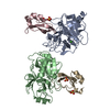
| ||||||||
|---|---|---|---|---|---|---|---|---|---|
| 1 | 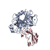
| ||||||||
| 2 | 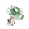
| ||||||||
| Unit cell |
|
-Components
| #1: Protein | Mass: 24088.062 Da / Num. of mol.: 2 / Fragment: UNP residues 24-247 / Mutation: K97D, R117H, S195A Source method: isolated from a genetically manipulated source Source: (gene. exp.) Homo sapiens (human) / Gene: PRSS1, TRP1, TRY1, TRYP1 / Organ: pancreas / Plasmid: pTRAP-T7 / Production host:  #2: Protein | Mass: 6342.387 Da / Num. of mol.: 2 / Fragment: UNP residues 36-90 Source method: isolated from a genetically manipulated source Source: (gene. exp.)  Komagataella pastoris (fungus) / Strain (production host): X-33 / References: UniProt: P00974 Komagataella pastoris (fungus) / Strain (production host): X-33 / References: UniProt: P00974#3: Chemical |   Mass: 40.078 Da / Num. of mol.: 2 / Source method: obtained synthetically / Formula: Ca Mass: 40.078 Da / Num. of mol.: 2 / Source method: obtained synthetically / Formula: Ca#4: Chemical | ChemComp-SO4 /   Mass: 96.063 Da / Num. of mol.: 4 / Source method: obtained synthetically / Formula: SO4 Mass: 96.063 Da / Num. of mol.: 4 / Source method: obtained synthetically / Formula: SO4#5: Water | ChemComp-HOH / |  Mass: 18.015 Da / Num. of mol.: 75 / Source method: isolated from a natural source / Formula: H2O Mass: 18.015 Da / Num. of mol.: 75 / Source method: isolated from a natural source / Formula: H2OHas protein modification | Y | |
|---|
-Experimental details
-Experiment
| Experiment | Method: X-RAY DIFFRACTION / Number of used crystals: 1 |
|---|
- Sample preparation
| Crystal | Density Matthews: 2.28 Å3/Da / Density % sol: 45.97 % |
|---|---|
| Crystal grow | Temperature: 298 K / Method: vapor diffusion, hanging drop / pH: 6.5 Details: 0.2M ammonium sulfate, 0.1 sodium cacodylate trihydrate, 30% PEG-8000 |
-Data collection
| Diffraction | Mean temperature: 100 K |
|---|---|
| Diffraction source | Source: SYNCHROTRON / Site: NSLS / Beamline: X29A / Wavelength: 0.979 Å |
| Detector | Type: ADSC QUANTUM 1 / Detector: CCD / Date: Feb 7, 2012 |
| Radiation | Protocol: SINGLE WAVELENGTH / Monochromatic (M) / Laue (L): M / Scattering type: x-ray |
| Radiation wavelength | Wavelength: 0.979 Å / Relative weight: 1 |
| Reflection | Resolution: 2.1→38.1 Å / Num. obs: 32877 / % possible obs: 98.1 % / Redundancy: 5.5 % / Rmerge(I) obs: 0.077 / Net I/σ(I): 15.9 |
| Reflection shell | Highest resolution: 2.1 Å / Redundancy: 4.1 % / Rmerge(I) obs: 0.256 / Mean I/σ(I) obs: 4.24 / % possible all: 85.3 |
- Processing
| Software |
| ||||||||||||||||||||||||||||||||||||||||||||||||||||||||||||
|---|---|---|---|---|---|---|---|---|---|---|---|---|---|---|---|---|---|---|---|---|---|---|---|---|---|---|---|---|---|---|---|---|---|---|---|---|---|---|---|---|---|---|---|---|---|---|---|---|---|---|---|---|---|---|---|---|---|---|---|---|---|
| Refinement | Method to determine structure: MOLECULAR REPLACEMENT Starting model: 2RA3 Resolution: 2.1→38.1 Å / Cor.coef. Fo:Fc: 0.935 / Cor.coef. Fo:Fc free: 0.898 / SU B: 5.945 / SU ML: 0.156 / Cross valid method: THROUGHOUT / σ(F): 0 / ESU R: 0.26 / ESU R Free: 0.225 / Stereochemistry target values: MAXIMUM LIKELIHOOD Details: HYDROGENS HAVE BEEN ADDED IN THE RIDING POSITIONS U VALUES : REFINED INDIVIDUALLY
| ||||||||||||||||||||||||||||||||||||||||||||||||||||||||||||
| Solvent computation | Ion probe radii: 0.8 Å / Shrinkage radii: 0.8 Å / VDW probe radii: 1.2 Å / Solvent model: MASK | ||||||||||||||||||||||||||||||||||||||||||||||||||||||||||||
| Displacement parameters | Biso max: 122.61 Å2 / Biso mean: 25.878 Å2 / Biso min: 12.62 Å2
| ||||||||||||||||||||||||||||||||||||||||||||||||||||||||||||
| Refinement step | Cycle: final / Resolution: 2.1→38.1 Å
| ||||||||||||||||||||||||||||||||||||||||||||||||||||||||||||
| Refine LS restraints |
| ||||||||||||||||||||||||||||||||||||||||||||||||||||||||||||
| LS refinement shell | Resolution: 2.099→2.153 Å / Total num. of bins used: 20
|
