 Movie
Movie Controller


+ Open data

- Basic information
| Entry | Database: PDB / ID: 1ldt | ||||||
|---|---|---|---|---|---|---|---|
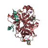
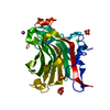
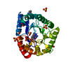
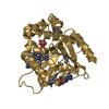
| Title | COMPLEX OF LEECH-DERIVED TRYPTASE INHIBITOR WITH PORCINE TRYPSIN | ||||||
 Components Components |
| ||||||
 Keywords Keywords | COMPLEX (HYDROLASE/INHIBITOR) / COMPLEX (HYDROLASE-INHIBITOR) / HYDROLASE / INHIBITOR / INFLAMMATION / TRYPTASE / COMPLEX (HYDROLASE-INHIBITOR) complex | ||||||
| Function / homology |  Function and homology information Function and homology informationtrypsin / digestion / serine-type endopeptidase inhibitor activity / serine-type endopeptidase activity / proteolysis / : / metal ion binding Similarity search - Function | ||||||
| Biological species |  Hirudo medicinalis (medicinal leech) Hirudo medicinalis (medicinal leech) | ||||||
| Method |  X-RAY DIFFRACTION / MOLECULAR REPLACEMENT / Resolution: 1.9 Å X-RAY DIFFRACTION / MOLECULAR REPLACEMENT / Resolution: 1.9 Å | ||||||
 Authors Authors | Stubbs, M.T. | ||||||
 Citation Citation | Journal: J.Biol.Chem. / Year: 1997 Title: The three-dimensional structure of recombinant leech-derived tryptase inhibitor in complex with trypsin. Implications for the structure of human mast cell tryptase and its inhibition. Authors: Stubbs, M.T. / Morenweiser, R. / Sturzebecher, J. / Bauer, M. / Bode, W. / Huber, R. / Piechottka, G.P. / Matschiner, G. / Sommerhoff, C.P. / Fritz, H. / Auerswald, E.A. #1: Journal: FEBS Lett. / Year: 1994Title: Structure of Leech Derived Tryptase Inhibitor (Ldti-C) in Solution Authors: Muhlhahn, P. / Czisch, M. / Morenweiser, R. / Habermann, B. / Engh, R.A. / Sommerhoff, C.P. / Auerswald, E.A. / Holak, T.A. #2: Journal: Biol.Chem.Hoppe-Seyler / Year: 1994Title: Recombinant Leech-Derived Tryptase Inhibitor: Construction, Production, Protein Chemical Characterization and Inhibition of HIV-1 Replication Authors: Auerswald, E.A. / Morenweiser, R. / Sommerhoff, C.P. / Piechottka, G.P. / Eckerskorn, C. / Gurtler, L.G. / Fritz, H. #3: Journal: Biol.Chem.Hoppe-Seyler / Year: 1994Title: A Kazal-Type Inhibitor of Human Mast Cell Tryptase: Isolation from the Medical Leech Hirudo Medicinalis, Characterization, and Sequence Analysis Authors: Sommerhoff, C.P. / Sollner, C. / Mentele, R. / Piechottka, G.P. / Auerswald, E.A. / Fritz, H. | ||||||
| History |
|
- Structure visualization
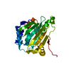
| Structure viewer | Molecule: MolmilJmol/JSmol |
|---|
- Downloads & links
-Download
| PDBx/mmCIF format | 1ldt.cif.gz | 67.8 KB | Display | PDBx/mmCIF format |
|---|---|---|---|---|
| PDB format | pdb1ldt.ent.gz | 48.8 KB | Display | PDB format |
| PDBx/mmJSON format | 1ldt.json.gz | Tree view | PDBx/mmJSON format | |
| Others |  Other downloads Other downloads |
-Validation report
| Arichive directory | https://data.pdbj.org/pub/pdb/validation_reports/ld/1ldtftp://data.pdbj.org/pub/pdb/validation_reports/ld/1ldt | HTTPS FTP |
|---|
-Related structure data
| Similar structure data |
|---|

-Links
 PDBj
PDBj

- Assembly
| Deposited unit | 
| ||||||||
|---|---|---|---|---|---|---|---|---|---|
| 1 |
| ||||||||
| Unit cell |
| ||||||||
| Components on special symmetry positions |
|

-Components
| #1: Protein | Mass: 23493.496 Da / Num. of mol.: 1 / Source method: isolated from a natural source / Source: (natural) |
|---|---|
| #2: Protein/peptide | Mass: 4750.614 Da / Num. of mol.: 1 Source method: isolated from a genetically manipulated source Details: LEECH-DERIVED TRYPTASE INHIBITOR / Source: (gene. exp.) Hirudo medicinalis (medicinal leech) / Production host:  |
| #3: Chemical | ChemComp-CA /   Mass: 40.078 Da / Num. of mol.: 1 / Source method: obtained synthetically / Formula: Ca Mass: 40.078 Da / Num. of mol.: 1 / Source method: obtained synthetically / Formula: Ca |
| #4: Water | ChemComp-HOH /  Mass: 18.015 Da / Num. of mol.: 149 / Source method: isolated from a natural source / Formula: H2O Mass: 18.015 Da / Num. of mol.: 149 / Source method: isolated from a natural source / Formula: H2O |
| Compound details | THIS STRUCTURE OF LDTI IN COMPLEX WITH TRYPSIN REVEALS STRUCTURAL ASPECTS OF THE MAST CELL ...THIS STRUCTURE OF LDTI IN COMPLEX WITH TRYPSIN REVEALS STRUCTURAL |
| Has protein modification | Y |
-Experimental details
-Experiment
| Experiment | Method: X-RAY DIFFRACTION / Number of used crystals: 1 |
|---|
- Sample preparation
| Crystal | Density Matthews: 2.33 Å3/Da / Density % sol: 47.28 % | ||||||||||||||||||||||||||||||||||||
|---|---|---|---|---|---|---|---|---|---|---|---|---|---|---|---|---|---|---|---|---|---|---|---|---|---|---|---|---|---|---|---|---|---|---|---|---|---|
| Crystal grow | pH: 8 / Details: 10% PEG 6000, 2.3M PHOSPHATE, PH 8.0 | ||||||||||||||||||||||||||||||||||||
| Crystal grow | *PLUS Method: vapor diffusion | ||||||||||||||||||||||||||||||||||||
| Components of the solutions | *PLUS
|
-Data collection
| Diffraction | Mean temperature: 287 K |
|---|---|
| Diffraction source | Source: ROTATING ANODE / Type: RIGAKU / Wavelength: 1.5418 |
| Detector | Type: MARRESEARCH / Detector: IMAGE PLATE / Date: Jan 1, 1995 / Details: MIRRORS |
| Radiation | Monochromator: NI FILTER / Monochromatic (M) / Laue (L): M / Scattering type: x-ray |
| Radiation wavelength | Wavelength: 1.5418 Å / Relative weight: 1 |
| Reflection | Resolution: 1.9→20 Å / Num. obs: 21466 / % possible obs: 98.6 % / Observed criterion σ(I): 2 / Redundancy: 6.5 % / Rmerge(I) obs: 0.082 / Rsym value: 0.082 / Net I/σ(I): 9 |
| Reflection shell | Resolution: 1.9→2 Å / Redundancy: 3.2 % / Rmerge(I) obs: 0.2 / Mean I/σ(I) obs: 4 / Rsym value: 0.2 / % possible all: 87.6 |
| Reflection | *PLUS Num. measured all: 139563 |
| Reflection shell | *PLUS % possible obs: 87.6 % |
- Processing
| Software |
| ||||||||||||||||||||||||||||||||||||||||||||||||||||||||||||
|---|---|---|---|---|---|---|---|---|---|---|---|---|---|---|---|---|---|---|---|---|---|---|---|---|---|---|---|---|---|---|---|---|---|---|---|---|---|---|---|---|---|---|---|---|---|---|---|---|---|---|---|---|---|---|---|---|---|---|---|---|---|
| Refinement | Method to determine structure: MOLECULAR REPLACEMENT Starting model: PORCINE TRYPSIN MODEL FROM TRYPSIN:MUNG BEAN INHIBITOR (LIN ET AL., EUR. J. BIOCHEM. 212, 549-555 (1993) Resolution: 1.9→6 Å / Data cutoff high absF: 10000000 / Data cutoff low absF: 0.001 / σ(F): 0 Details: THE LDTI MOIETY IS WELL DEFINED IN THE VICINITY OF THE PROTEINASE, BUT IS CHARACTERIZED BY ELEVATED TEMPERATURE FACTORS AND DISRUPTED DENSITY FURTHER AWAY FROM TRYPSIN. IN PARTICULAR, AMINO ...Details: THE LDTI MOIETY IS WELL DEFINED IN THE VICINITY OF THE PROTEINASE, BUT IS CHARACTERIZED BY ELEVATED TEMPERATURE FACTORS AND DISRUPTED DENSITY FURTHER AWAY FROM TRYPSIN. IN PARTICULAR, AMINO ACID RESIDUES LYS L 1I - LYS L 2I, GLY L 15 I - ARG L 19I, SER L 33I - SER L 36I AND THE C-TERMINAL RESIDUES PRO L 41I - ASN L 46I ARE DEFINED BY EITHER WEAK OR NO ELECTRON DENSITY. ACCORDINGLY, THE COORDINATES FOR PRO L 41I - ASN L 46I ARE NOT RELIABLE.
| ||||||||||||||||||||||||||||||||||||||||||||||||||||||||||||
| Refine analyze | Luzzati d res low obs: 6 Å | ||||||||||||||||||||||||||||||||||||||||||||||||||||||||||||
| Refinement step | Cycle: LAST / Resolution: 1.9→6 Å
| ||||||||||||||||||||||||||||||||||||||||||||||||||||||||||||
| Refine LS restraints |
| ||||||||||||||||||||||||||||||||||||||||||||||||||||||||||||
| LS refinement shell | Resolution: 1.9→1.93 Å / Total num. of bins used: 20
| ||||||||||||||||||||||||||||||||||||||||||||||||||||||||||||
| Xplor file |
| ||||||||||||||||||||||||||||||||||||||||||||||||||||||||||||
| Software | *PLUS Name: X-PLOR / Version: 3.1 / Classification: refinement | ||||||||||||||||||||||||||||||||||||||||||||||||||||||||||||
| Refinement | *PLUS | ||||||||||||||||||||||||||||||||||||||||||||||||||||||||||||
| Solvent computation | *PLUS | ||||||||||||||||||||||||||||||||||||||||||||||||||||||||||||
| Displacement parameters | *PLUS Biso mean: 21 Å2 | ||||||||||||||||||||||||||||||||||||||||||||||||||||||||||||
| Refine LS restraints | *PLUS
| ||||||||||||||||||||||||||||||||||||||||||||||||||||||||||||
| LS refinement shell | *PLUS Rfactor obs: 0.29 |
