 Movie
Movie Controller

[English] 日本語
 Yorodumi
- PDB-1z9d: Crystal structure of a putative uridylate kinase (UMP-kinase) fro... -
+ Open data

- Basic information
| Entry | Database: PDB / ID: 1z9d | ||||||
|---|---|---|---|---|---|---|---|
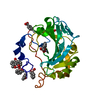
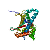
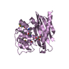
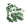
| Title | Crystal structure of a putative uridylate kinase (UMP-kinase) from Streptococcus pyogenes | ||||||
 Components Components | uridylate kinase | ||||||
 Keywords Keywords | TRANSFERASE / Structural Genomics / Protein Structure Initiative / NYSGXRC / T1668 / pyrH / putative uridylate kinase / UMP-kinase / PSI / New York SGX Research Center for Structural Genomics | ||||||
| Function / homology |  Function and homology information Function and homology informationUMP kinase / UMP kinase activity / 'de novo' CTP biosynthetic process / UDP biosynthetic process / ATP binding / cytoplasm Similarity search - Function | ||||||
| Biological species |  Streptococcus pyogenes (bacteria) Streptococcus pyogenes (bacteria) | ||||||
| Method |  X-RAY DIFFRACTION / SYNCHROTRON / SAD aided by molecular replacement / Resolution: 2.8 Å X-RAY DIFFRACTION / SYNCHROTRON / SAD aided by molecular replacement / Resolution: 2.8 Å | ||||||
 Authors Authors | Rajashankar, K.R. / Kniewel, R. / Lee, K. / Lima, C.D. / Burley, S.K. / New York SGX Research Center for Structural Genomics (NYSGXRC) | ||||||
 Citation Citation | Journal: To be Published Title: Crystal structure of a putative uridylate kinase (UMP-kinase) from Streptococcus pyogenes Authors: Rajashankar, K.R. / Kniewel, R. / Lee, K. / Lima, C.D. | ||||||
| History |
|
- Structure visualization
| Structure viewer | Molecule: MolmilJmol/JSmol |
|---|
- Downloads & links
-Download
| PDBx/mmCIF format | 1z9d.cif.gz | 146.1 KB | Display | PDBx/mmCIF format |
|---|---|---|---|---|
| PDB format | pdb1z9d.ent.gz | 116.1 KB | Display | PDB format |
| PDBx/mmJSON format | 1z9d.json.gz | Tree view | PDBx/mmJSON format | |
| Others |  Other downloads Other downloads |
-Validation report
| Arichive directory | https://data.pdbj.org/pub/pdb/validation_reports/z9/1z9dftp://data.pdbj.org/pub/pdb/validation_reports/z9/1z9d | HTTPS FTP |
|---|
-Related structure data
| Similar structure data | |
|---|---|
| Other databases |


-Links
 PDBj
PDBj- Assembly
| Deposited unit | 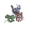
| ||||||||||
|---|---|---|---|---|---|---|---|---|---|---|---|
| 1 | 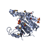
| ||||||||||
| 2 | 
| ||||||||||
| 3 | 
| ||||||||||
| 4 | 
| ||||||||||
| Unit cell |
| ||||||||||
| Details | UMP-Kinase from B. subtilis has a sequence identity of 40% to T1668 and is known to exist as a hexamer (a trimer of dimers). This fact has been experimentally tested via Gel filtration(C. Gagyi et. al. Eur. J. Biochem. 270, 3196-3204). However biologically active species are monomers. The hexamer can be generated by symmetry operation -X+1,-Y,Z. |
-Components
| #1: Protein | Mass: 27607.812 Da / Num. of mol.: 3 Source method: isolated from a genetically manipulated source Source: (gene. exp.) Streptococcus pyogenes (bacteria) / Gene: pyrH / Plasmid: PET T7 / Production host: References: UniProt: P65938, Transferases; Transferring phosphorus-containing groups; Phosphotransferases with a phosphate group as acceptor #2: Chemical | ChemComp-SO4 /   Mass: 96.063 Da / Num. of mol.: 9 / Source method: obtained synthetically / Formula: SO4 Mass: 96.063 Da / Num. of mol.: 9 / Source method: obtained synthetically / Formula: SO4#3: Water | ChemComp-HOH / |  Mass: 18.015 Da / Num. of mol.: 88 / Source method: isolated from a natural source / Formula: H2O Mass: 18.015 Da / Num. of mol.: 88 / Source method: isolated from a natural source / Formula: H2OHas protein modification | Y | |
|---|
-Experimental details
-Experiment
| Experiment | Method: X-RAY DIFFRACTION / Number of used crystals: 1 |
|---|
- Sample preparation
| Crystal | Density Matthews: 2.68 Å3/Da / Density % sol: 53.72 % |
|---|---|
| Crystal grow | Temperature: 291 K / Method: vapor diffusion, hanging drop / pH: 8.5 Details: 2M Ammonium sulfate, 0.15M Tris pH 8.5, VAPOR DIFFUSION, HANGING DROP, temperature 291K |
-Data collection
| Diffraction | Mean temperature: 100 K |
|---|---|
| Diffraction source | Source: SYNCHROTRON / Site: APS  / Beamline: 31-ID / Wavelength: 0.98 Å / Beamline: 31-ID / Wavelength: 0.98 Å |
| Detector | Type: MARRESEARCH / Detector: CCD / Date: Jun 26, 2004 / Details: Diamond monochromator and downstream mirror |
| Radiation | Monochromator: Flat Diamond 111 / Protocol: SINGLE WAVELENGTH / Monochromatic (M) / Laue (L): M / Scattering type: x-ray |
| Radiation wavelength | Wavelength: 0.98 Å / Relative weight: 1 |
| Reflection | Resolution: 2.8→20 Å / Num. all: 38306 / Num. obs: 38306 / % possible obs: 93.1 % / Observed criterion σ(I): -3 / Redundancy: 11.04 % / Biso Wilson estimate: 59.5 Å2 / Rsym value: 0.08 |
| Reflection shell | Resolution: 2.8→2.9 Å / Mean I/σ(I) obs: 2.05 / Num. unique all: 3849 / Rsym value: 0.382 / % possible all: 92.9 |
- Processing
| Software |
| ||||||||||||||||||||||||||||||||||||
|---|---|---|---|---|---|---|---|---|---|---|---|---|---|---|---|---|---|---|---|---|---|---|---|---|---|---|---|---|---|---|---|---|---|---|---|---|---|
| Refinement | Method to determine structure: SAD aided by molecular replacement Resolution: 2.8→19.72 Å / Rfactor Rfree error: 0.006 / Data cutoff high absF: 222033.22 / Data cutoff low absF: 0 / Isotropic thermal model: RESTRAINED / Cross valid method: THROUGHOUT / σ(F): 0 / Stereochemistry target values: Engh & Huber Details: A molecular replacement solution was obtained using a model derived from pdb entry 1YBD. MR phases were used to locate Se sites. Experimental phases were calculated using a Se-substructure ...Details: A molecular replacement solution was obtained using a model derived from pdb entry 1YBD. MR phases were used to locate Se sites. Experimental phases were calculated using a Se-substructure containing 26 Se sites. Nine sulfate groups were located. Sulfates D4 - D9 mimic phosphate group of ATP at the ATP binding pocket.
| ||||||||||||||||||||||||||||||||||||
| Solvent computation | Solvent model: FLAT MODEL / Bsol: 11.7658 Å2 / ksol: 0.318634 e/Å3 | ||||||||||||||||||||||||||||||||||||
| Displacement parameters | Biso mean: 41.5 Å2
| ||||||||||||||||||||||||||||||||||||
| Refine analyze |
| ||||||||||||||||||||||||||||||||||||
| Refinement step | Cycle: LAST / Resolution: 2.8→19.72 Å
| ||||||||||||||||||||||||||||||||||||
| Refine LS restraints |
| ||||||||||||||||||||||||||||||||||||
| LS refinement shell | Resolution: 2.8→2.97 Å / Rfactor Rfree error: 0.024 / Total num. of bins used: 6
| ||||||||||||||||||||||||||||||||||||
| Xplor file |
|
