 Movie
Movie Controller

[English] 日本語
 Yorodumi
- PDB-4xcl: N-terminal domain of Hsp90 from Dictyostelium discoideum in compl... -
+ Open data

- Basic information
| Entry | Database: PDB / ID: 4xcl | ||||||
|---|---|---|---|---|---|---|---|
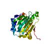
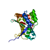
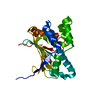
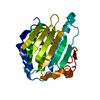
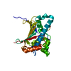
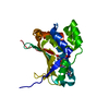
| Title | N-terminal domain of Hsp90 from Dictyostelium discoideum in complex with AGS | ||||||
 Components Components | Heat shock cognate 90 kDa protein | ||||||
 Keywords Keywords | CHAPERONE / Hsp90 / AGS | ||||||
| Function / homology |  Function and homology information Function and homology informationregulation of aggregation involved in sorocarp development / Tetrahydrobiopterin (BH4) synthesis, recycling, salvage and regulation / eNOS activation / HSP90 chaperone cycle for steroid hormone receptors (SHR) in the presence of ligand / HSF1 activation / HSF1-dependent transactivation / Sema3A PAK dependent Axon repulsion / VEGFR2 mediated vascular permeability / The NLRP3 inflammasome / : ...regulation of aggregation involved in sorocarp development / Tetrahydrobiopterin (BH4) synthesis, recycling, salvage and regulation / eNOS activation / HSP90 chaperone cycle for steroid hormone receptors (SHR) in the presence of ligand / HSF1 activation / HSF1-dependent transactivation / Sema3A PAK dependent Axon repulsion / VEGFR2 mediated vascular permeability / The NLRP3 inflammasome / : / Extra-nuclear estrogen signaling / Regulation of actin dynamics for phagocytic cup formation / Neutrophil degranulation / phagocytic vesicle / ATP-dependent protein folding chaperone / : / cellular response to heat / extracellular matrix / protein folding / protein stabilization / perinuclear region of cytoplasm / ATP hydrolysis activity / protein-containing complex / ATP binding / plasma membrane / cytosol Similarity search - Function | ||||||
| Biological species |  | ||||||
| Method |  X-RAY DIFFRACTION / SYNCHROTRON / MOLECULAR REPLACEMENT / Resolution: 1.21 Å X-RAY DIFFRACTION / SYNCHROTRON / MOLECULAR REPLACEMENT / Resolution: 1.21 Å | ||||||
 Authors Authors | Raman, S. / Suguna, K. | ||||||
 Citation Citation | Journal: Sci Rep / Year: 2015 Title: First Structural View of a Peptide Interacting with the Nucleotide Binding Domain of Heat Shock Protein 90 Authors: Raman, S. / Singh, M. / Tatu, U. / Suguna, K. | ||||||
| History |
|
- Structure visualization
| Structure viewer | Molecule: MolmilJmol/JSmol |
|---|
- Downloads & links
-Download
| PDBx/mmCIF format | 4xcl.cif.gz | 119 KB | Display | PDBx/mmCIF format |
|---|---|---|---|---|
| PDB format | pdb4xcl.ent.gz | 89.1 KB | Display | PDB format |
| PDBx/mmJSON format | 4xcl.json.gz | Tree view | PDBx/mmJSON format | |
| Others |  Other downloads Other downloads |
-Validation report
| Arichive directory | https://data.pdbj.org/pub/pdb/validation_reports/xc/4xclftp://data.pdbj.org/pub/pdb/validation_reports/xc/4xcl | HTTPS FTP |
|---|
-Related structure data
| Related structure data |  4xc0C  4xcjSC  4xd8C  4xdmC  4xe2C  4xkaC 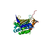 4xkoC C: citing same article ( S: Starting model for refinement |
|---|---|
| Similar structure data |


-Links
 PDBj
PDBj


- Assembly
| Deposited unit | 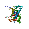
| ||||||||
|---|---|---|---|---|---|---|---|---|---|
| 1 |
| ||||||||
| Unit cell |
|
-Components
| #1: Protein | Mass: 29226.871 Da / Num. of mol.: 1 / Fragment: N-terminal domain, UNP residues 1-223 Source method: isolated from a genetically manipulated source Source: (gene. exp.)  |
|---|---|
| #2: Chemical | ChemComp-AGS /   Mass: 523.247 Da / Num. of mol.: 1 / Source method: obtained synthetically / Formula: C10H16N5O12P3S / Comment: ATP-gamma-S, energy-carrying molecule analogue*YM Mass: 523.247 Da / Num. of mol.: 1 / Source method: obtained synthetically / Formula: C10H16N5O12P3S / Comment: ATP-gamma-S, energy-carrying molecule analogue*YM |
| #3: Chemical | ChemComp-MG /   Mass: 24.305 Da / Num. of mol.: 1 / Source method: obtained synthetically / Formula: Mg Mass: 24.305 Da / Num. of mol.: 1 / Source method: obtained synthetically / Formula: Mg |
| #4: Water | ChemComp-HOH /  Mass: 18.015 Da / Num. of mol.: 288 / Source method: isolated from a natural source / Formula: H2O Mass: 18.015 Da / Num. of mol.: 288 / Source method: isolated from a natural source / Formula: H2O |
-Experimental details
-Experiment
| Experiment | Method: X-RAY DIFFRACTION |
|---|
- Sample preparation
| Crystal | Density Matthews: 1.91 Å3/Da / Density % sol: 35.5 % |
|---|---|
| Crystal grow | Temperature: 290 K / Method: vapor diffusion, hanging drop / pH: 7.5 / Details: 0.1M Hepes, PEG 3350 / PH range: 7.0-8.5 |
-Data collection
| Diffraction | Mean temperature: 100 K |
|---|---|
| Diffraction source | Source: SYNCHROTRON / Site: ESRF  / Beamline: BM14 / Wavelength: 0.97625 Å / Beamline: BM14 / Wavelength: 0.97625 Å |
| Detector | Type: MARMOSAIC 225 mm CCD / Detector: CCD / Date: Jul 4, 2013 |
| Radiation | Monochromator: Si(111) / Protocol: SINGLE WAVELENGTH / Monochromatic (M) / Laue (L): M / Scattering type: x-ray |
| Radiation wavelength | Wavelength: 0.97625 Å / Relative weight: 1 |
| Reflection | Resolution: 1.21→56.94 Å / Num. obs: 64120 / % possible obs: 95.7 % / Redundancy: 9.1 % / Rmerge(I) obs: 0.06 / Net I/σ(I): 18.6 |
| Reflection shell | Resolution: 1.21→1.28 Å / Redundancy: 7.4 % / Rmerge(I) obs: 0.369 / Mean I/σ(I) obs: 5.2 / % possible all: 92.5 |
- Processing
| Software | Name: REFMAC / Version: 5.5.0109 / Classification: refinement | ||||||||||||||||||||||||||||||||||||||||||||||||||||||||||||||||||||||||||||||||||||||||||||||||||||||||||||||||||||||||||||||||||||||||||||||||||||||||||||||||||||||||||||||||||||||
|---|---|---|---|---|---|---|---|---|---|---|---|---|---|---|---|---|---|---|---|---|---|---|---|---|---|---|---|---|---|---|---|---|---|---|---|---|---|---|---|---|---|---|---|---|---|---|---|---|---|---|---|---|---|---|---|---|---|---|---|---|---|---|---|---|---|---|---|---|---|---|---|---|---|---|---|---|---|---|---|---|---|---|---|---|---|---|---|---|---|---|---|---|---|---|---|---|---|---|---|---|---|---|---|---|---|---|---|---|---|---|---|---|---|---|---|---|---|---|---|---|---|---|---|---|---|---|---|---|---|---|---|---|---|---|---|---|---|---|---|---|---|---|---|---|---|---|---|---|---|---|---|---|---|---|---|---|---|---|---|---|---|---|---|---|---|---|---|---|---|---|---|---|---|---|---|---|---|---|---|---|---|---|---|
| Refinement | Method to determine structure: MOLECULAR REPLACEMENT Starting model: 4XCJ Resolution: 1.21→56.94 Å / Cor.coef. Fo:Fc: 0.974 / Cor.coef. Fo:Fc free: 0.966 / SU B: 1.11 / SU ML: 0.023 / Cross valid method: FREE R-VALUE / ESU R: 0.045 / ESU R Free: 0.042 / Stereochemistry target values: MAXIMUM LIKELIHOOD / Details: HYDROGENS HAVE BEEN ADDED IN THE RIDING POSITIONS
| ||||||||||||||||||||||||||||||||||||||||||||||||||||||||||||||||||||||||||||||||||||||||||||||||||||||||||||||||||||||||||||||||||||||||||||||||||||||||||||||||||||||||||||||||||||||
| Solvent computation | Ion probe radii: 0.8 Å / Shrinkage radii: 0.8 Å / VDW probe radii: 1.4 Å / Solvent model: MASK | ||||||||||||||||||||||||||||||||||||||||||||||||||||||||||||||||||||||||||||||||||||||||||||||||||||||||||||||||||||||||||||||||||||||||||||||||||||||||||||||||||||||||||||||||||||||
| Displacement parameters | Biso mean: 18.067 Å2
| ||||||||||||||||||||||||||||||||||||||||||||||||||||||||||||||||||||||||||||||||||||||||||||||||||||||||||||||||||||||||||||||||||||||||||||||||||||||||||||||||||||||||||||||||||||||
| Refinement step | Cycle: LAST / Resolution: 1.21→56.94 Å
| ||||||||||||||||||||||||||||||||||||||||||||||||||||||||||||||||||||||||||||||||||||||||||||||||||||||||||||||||||||||||||||||||||||||||||||||||||||||||||||||||||||||||||||||||||||||
| Refine LS restraints |
|