 Movie
Movie Controller

[English] 日本語
 Yorodumi
- PDB-6zqt: Crystal structure of the RLIP76 Ral binding domain mutant (E427H/... -
+ Open data

- Basic information
| Entry | Database: PDB / ID: 6zqt | ||||||
|---|---|---|---|---|---|---|---|
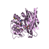
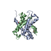
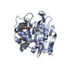
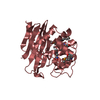
| Title | Crystal structure of the RLIP76 Ral binding domain mutant (E427H/Q433L/K440R) in complex with RalB-GMPPNP | ||||||
 Components Components |
| ||||||
 Keywords Keywords | PROTEIN BINDING / RalB / RLIP76 / Ral binding domain / coiled-coil / small GTPase / G protein | ||||||
| Function / homology |  Function and homology information Function and homology informationdoxorubicin transport / regulation of exocyst assembly / regulation of exocyst localization / ABC-type glutathione-S-conjugate transporter / ABC-type glutathione S-conjugate transporter activity / positive regulation of autophagosome assembly / regulation of GTPase activity / xenobiotic detoxification by transmembrane export across the plasma membrane / positive regulation of innate immune response / regulation of small GTPase mediated signal transduction ...doxorubicin transport / regulation of exocyst assembly / regulation of exocyst localization / ABC-type glutathione-S-conjugate transporter / ABC-type glutathione S-conjugate transporter activity / positive regulation of autophagosome assembly / regulation of GTPase activity / xenobiotic detoxification by transmembrane export across the plasma membrane / positive regulation of innate immune response / regulation of small GTPase mediated signal transduction / regulation of Cdc42 protein signal transduction / ABC-type xenobiotic transporter / ABC-type xenobiotic transporter activity / positive regulation of mitochondrial fission / small GTPase-mediated signal transduction / CDC42 GTPase cycle / ATPase-coupled transmembrane transporter activity / positive regulation of GTPase activity / xenobiotic transmembrane transporter activity / transmembrane transporter activity / positive regulation of epidermal growth factor receptor signaling pathway / p38MAPK events / RAC1 GTPase cycle / receptor-mediated endocytosis / GTPase activator activity / small monomeric GTPase / positive regulation of protein phosphorylation / receptor internalization / transmembrane transport / small GTPase binding / chemotaxis / spindle pole / GDP binding / ATPase binding / G protein activity / midbody / Ras protein signal transduction / nuclear body / cell division / GTPase activity / apoptotic process / ubiquitin protein ligase binding / GTP binding / signal transduction / mitochondrion / extracellular exosome / nucleoplasm / ATP binding / membrane / plasma membrane / cytosol Similarity search - Function | ||||||
| Biological species |  Homo sapiens (human) Homo sapiens (human) | ||||||
| Method |  X-RAY DIFFRACTION / SYNCHROTRON / MOLECULAR REPLACEMENT / molecular replacement / Resolution: 1.51 Å X-RAY DIFFRACTION / SYNCHROTRON / MOLECULAR REPLACEMENT / molecular replacement / Resolution: 1.51 Å | ||||||
 Authors Authors | Hurd, C. / Brear, P. / Revell, J. / Ross, S. / Mott, H. / Owen, D. | ||||||
 Citation Citation | Journal: J.Biol.Chem. / Year: 2020 Title: Affinity maturation of the RLIP76 Ral binding domain to inform the design of stapled peptides targeting the Ral GTPases. Authors: Hurd, C.A. / Brear, P. / Revell, J. / Ross, S. / Mott, H.R. / Owen, D. | ||||||
| History |
|
- Structure visualization
| Structure viewer | Molecule: MolmilJmol/JSmol |
|---|
- Downloads & links
-Download
| PDBx/mmCIF format | 6zqt.cif.gz | 117.1 KB | Display | PDBx/mmCIF format |
|---|---|---|---|---|
| PDB format | pdb6zqt.ent.gz | 87.9 KB | Display | PDB format |
| PDBx/mmJSON format | 6zqt.json.gz | Tree view | PDBx/mmJSON format | |
| Others |  Other downloads Other downloads |
-Validation report
| Arichive directory | https://data.pdbj.org/pub/pdb/validation_reports/zq/6zqtftp://data.pdbj.org/pub/pdb/validation_reports/zq/6zqt | HTTPS FTP |
|---|
-Related structure data
| Related structure data |  6zrnC  2kwiS S: Starting model for refinement C: citing same article ( |
|---|---|
| Similar structure data |


-Links
 PDBj
PDBj


- Assembly
| Deposited unit | 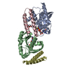
| ||||||||
|---|---|---|---|---|---|---|---|---|---|
| 1 | 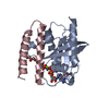
| ||||||||
| 2 | 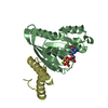
| ||||||||
| Unit cell |
|
-Components
-Protein , 2 types, 4 molecules ABCD
| #1: Protein | Mass: 20998.691 Da / Num. of mol.: 2 Source method: isolated from a genetically manipulated source Source: (gene. exp.) Homo sapiens (human) / Gene: RALB / Production host:  #2: Protein | Mass: 6889.968 Da / Num. of mol.: 2 Source method: isolated from a genetically manipulated source Source: (gene. exp.) Homo sapiens (human) / Gene: RALBP1, RLIP1, RLIP76 / Production host: |
|---|
-Non-polymers , 4 types, 366 molecules 


| #3: Chemical |  Mass: 522.196 Da / Num. of mol.: 2 / Source method: obtained synthetically / Formula: C10H17N6O13P3 Mass: 522.196 Da / Num. of mol.: 2 / Source method: obtained synthetically / Formula: C10H17N6O13P3Comment: GppNHp, GMPPNP, energy-carrying molecule analogue*YM #4: Chemical |  Mass: 24.305 Da / Num. of mol.: 2 / Source method: obtained synthetically / Formula: Mg Mass: 24.305 Da / Num. of mol.: 2 / Source method: obtained synthetically / Formula: Mg#5: Chemical | ChemComp-GOL / |  Mass: 92.094 Da / Num. of mol.: 1 / Source method: obtained synthetically / Formula: C3H8O3 Mass: 92.094 Da / Num. of mol.: 1 / Source method: obtained synthetically / Formula: C3H8O3#6: Water | ChemComp-HOH / | Mass: 18.015 Da / Num. of mol.: 361 / Source method: isolated from a natural source / Formula: H2O |
|---|
-Details
| Has ligand of interest | N |
|---|
-Experimental details
-Experiment
| Experiment | Method: X-RAY DIFFRACTION / Number of used crystals: 1 |
|---|
- Sample preparation
| Crystal | Density Matthews: 2.16 Å3/Da / Density % sol: 43.04 % |
|---|---|
| Crystal grow | Temperature: 293 K / Method: vapor diffusion, sitting drop / pH: 9 / Details: 0.1 M Bicine 9.0 pH, 30% w/v PEG 6000 |
-Data collection
| Diffraction | Mean temperature: 80 K / Serial crystal experiment: N | ||||||||||||||||||||||||||||||
|---|---|---|---|---|---|---|---|---|---|---|---|---|---|---|---|---|---|---|---|---|---|---|---|---|---|---|---|---|---|---|---|
| Diffraction source | Source: SYNCHROTRON / Site: Diamond  / Beamline: I04 / Wavelength: 0.9795 Å / Beamline: I04 / Wavelength: 0.9795 Å | ||||||||||||||||||||||||||||||
| Detector | Type: DECTRIS PILATUS 6M-F / Detector: PIXEL / Date: Aug 6, 2017 | ||||||||||||||||||||||||||||||
| Radiation | Protocol: SINGLE WAVELENGTH / Monochromatic (M) / Laue (L): M / Scattering type: x-ray | ||||||||||||||||||||||||||||||
| Radiation wavelength | Wavelength: 0.9795 Å / Relative weight: 1 | ||||||||||||||||||||||||||||||
| Reflection | Resolution: 1.51→50.404 Å / Num. obs: 75045 / % possible obs: 99.7 % / Redundancy: 7.6 % / CC1/2: 0.999 / Rmerge(I) obs: 0.061 / Rpim(I) all: 0.022 / Rrim(I) all: 0.065 / Net I/σ(I): 18.3 | ||||||||||||||||||||||||||||||
| Reflection shell | Diffraction-ID: 1
|
-Phasing
| Phasing | Method: molecular replacement |
|---|
- Processing
| Software |
| ||||||||||||||||||||||||||||||||||||||||||||||||||||||||||||||||||||||||||||||||||||||||||||||||||||||||||||||||||||||||||||||||||||||||||||||||||||||||||||||||||||||||
|---|---|---|---|---|---|---|---|---|---|---|---|---|---|---|---|---|---|---|---|---|---|---|---|---|---|---|---|---|---|---|---|---|---|---|---|---|---|---|---|---|---|---|---|---|---|---|---|---|---|---|---|---|---|---|---|---|---|---|---|---|---|---|---|---|---|---|---|---|---|---|---|---|---|---|---|---|---|---|---|---|---|---|---|---|---|---|---|---|---|---|---|---|---|---|---|---|---|---|---|---|---|---|---|---|---|---|---|---|---|---|---|---|---|---|---|---|---|---|---|---|---|---|---|---|---|---|---|---|---|---|---|---|---|---|---|---|---|---|---|---|---|---|---|---|---|---|---|---|---|---|---|---|---|---|---|---|---|---|---|---|---|---|---|---|---|---|---|---|---|
| Refinement | Method to determine structure: MOLECULAR REPLACEMENT Starting model: 2KWI Resolution: 1.51→50.404 Å / SU ML: 0.19 / Cross valid method: THROUGHOUT / σ(F): 1.34 / Phase error: 26.08 / Stereochemistry target values: ML
| ||||||||||||||||||||||||||||||||||||||||||||||||||||||||||||||||||||||||||||||||||||||||||||||||||||||||||||||||||||||||||||||||||||||||||||||||||||||||||||||||||||||||
| Solvent computation | Shrinkage radii: 0.9 Å / VDW probe radii: 1.11 Å / Solvent model: FLAT BULK SOLVENT MODEL | ||||||||||||||||||||||||||||||||||||||||||||||||||||||||||||||||||||||||||||||||||||||||||||||||||||||||||||||||||||||||||||||||||||||||||||||||||||||||||||||||||||||||
| Displacement parameters | Biso max: 80.78 Å2 / Biso mean: 34.4674 Å2 / Biso min: 17.13 Å2 | ||||||||||||||||||||||||||||||||||||||||||||||||||||||||||||||||||||||||||||||||||||||||||||||||||||||||||||||||||||||||||||||||||||||||||||||||||||||||||||||||||||||||
| Refinement step | Cycle: final / Resolution: 1.51→50.404 Å
| ||||||||||||||||||||||||||||||||||||||||||||||||||||||||||||||||||||||||||||||||||||||||||||||||||||||||||||||||||||||||||||||||||||||||||||||||||||||||||||||||||||||||
| LS refinement shell | Refine-ID: X-RAY DIFFRACTION / Rfactor Rfree error: 0
|
