 Movie
Movie Controller


+ Open data

- Basic information
| Entry | Database: PDB / ID: 4mit | ||||||
|---|---|---|---|---|---|---|---|
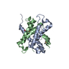
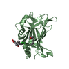
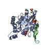
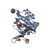
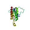
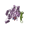
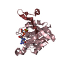
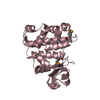
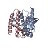
| Title | Crystal structure of E. histolytica RacC bound to the EhPAK4 PBD | ||||||
 Components Components |
| ||||||
 Keywords Keywords | SIGNALING PROTEIN / G domain / p21 binding domain / CRIB motif / hydrolase / kinase / GTP binding | ||||||
| Function / homology |  Function and homology information Function and homology informationhistone H4S1 kinase activity / histone H2AS121 kinase activity / histone H3S57 kinase activity / histone H2BS14 kinase activity / histone H3S28 kinase activity / histone H2AS1 kinase activity / histone H3T6 kinase activity / histone H2BS36 kinase activity / ribosomal protein S6 kinase activity / histone H2AT120 kinase activity ...histone H4S1 kinase activity / histone H2AS121 kinase activity / histone H3S57 kinase activity / histone H2BS14 kinase activity / histone H3S28 kinase activity / histone H2AS1 kinase activity / histone H3T6 kinase activity / histone H2BS36 kinase activity / ribosomal protein S6 kinase activity / histone H2AT120 kinase activity / Rho-dependent protein serine/threonine kinase activity / AMP-activated protein kinase activity / eukaryotic translation initiation factor 2alpha kinase activity / 3-phosphoinositide-dependent protein kinase activity / histone H3T3 kinase activity / histone H3T45 kinase activity / DNA-dependent protein kinase activity / histone H2AXS139 kinase activity / histone H3T11 kinase activity / histone H3S10 kinase activity / small GTPase-mediated signal transduction / small monomeric GTPase / cytoskeleton / GTPase activity / GTP binding / ATP binding / metal ion binding / plasma membrane / cytoplasm Similarity search - Function | ||||||
| Biological species |   Entamoeba histolytica (eukaryote) Entamoeba histolytica (eukaryote) | ||||||
| Method |  X-RAY DIFFRACTION / SYNCHROTRON / MOLECULAR REPLACEMENT / Resolution: 2.35 Å X-RAY DIFFRACTION / SYNCHROTRON / MOLECULAR REPLACEMENT / Resolution: 2.35 Å | ||||||
 Authors Authors | Bosch, D.E. / Siderovski, D.P. | ||||||
 Citation Citation | Journal: Biochemistry / Year: 2015 Title: Entamoeba histolytica RacC Selectively Engages p21-Activated Kinase Effectors. Authors: Bosch, D.E. / Siderovski, D.P. | ||||||
| History |
|
- Structure visualization
| Structure viewer | Molecule: MolmilJmol/JSmol |
|---|
- Downloads & links
-Download
| PDBx/mmCIF format | 4mit.cif.gz | 196.6 KB | Display | PDBx/mmCIF format |
|---|---|---|---|---|
| PDB format | pdb4mit.ent.gz | 156.1 KB | Display | PDB format |
| PDBx/mmJSON format | 4mit.json.gz | Tree view | PDBx/mmJSON format | |
| Others |  Other downloads Other downloads |
-Validation report
| Arichive directory | https://data.pdbj.org/pub/pdb/validation_reports/mi/4mitftp://data.pdbj.org/pub/pdb/validation_reports/mi/4mit | HTTPS FTP |
|---|
-Related structure data
| Related structure data | 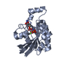 3th5S S: Starting model for refinement |
|---|---|
| Similar structure data |

-Links
 PDBj
PDBj


- Assembly
| Deposited unit | 
| ||||||||
|---|---|---|---|---|---|---|---|---|---|
| 1 | 
| ||||||||
| 2 | 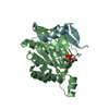
| ||||||||
| 3 | 
| ||||||||
| 4 | 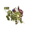
| ||||||||
| Unit cell |
|
-Components
| #1: Protein | Mass: 20645.807 Da / Num. of mol.: 4 / Mutation: Q65L Source method: isolated from a genetically manipulated source Source: (gene. exp.) Entamoeba histolytica (eukaryote) / Strain: HM-1:IMSS / Gene: EhRacC, KM1_331620 / Plasmid: pLIC His / Production host:  References: UniProt: M7WE85, UniProt: Q24816*PLUS, small monomeric GTPase #2: Protein | Mass: 7505.105 Da / Num. of mol.: 4 / Fragment: EhPAK4 PBD, unp residues 33-99 Source method: isolated from a genetically manipulated source Source: (gene. exp.) Entamoeba histolytica (eukaryote) / Strain: HM-1:IMSS / Gene: EHI7A_018700, EhPAK4 / Plasmid: pLIC His / Production host: #3: Chemical | ChemComp-GTP /   Mass: 523.180 Da / Num. of mol.: 4 / Source method: obtained synthetically / Formula: C10H16N5O14P3 / Comment: GTP, energy-carrying molecule*YM Mass: 523.180 Da / Num. of mol.: 4 / Source method: obtained synthetically / Formula: C10H16N5O14P3 / Comment: GTP, energy-carrying molecule*YM#4: Chemical | ChemComp-MG /   Mass: 24.305 Da / Num. of mol.: 12 / Source method: obtained synthetically / Formula: Mg Mass: 24.305 Da / Num. of mol.: 12 / Source method: obtained synthetically / Formula: Mg#5: Water | ChemComp-HOH / |  Mass: 18.015 Da / Num. of mol.: 341 / Source method: isolated from a natural source / Formula: H2O Mass: 18.015 Da / Num. of mol.: 341 / Source method: isolated from a natural source / Formula: H2O |
|---|
-Experimental details
-Experiment
| Experiment | Method: X-RAY DIFFRACTION / Number of used crystals: 1 |
|---|
- Sample preparation
| Crystal | Density Matthews: 2.25 Å3/Da / Density % sol: 45.4 % |
|---|---|
| Crystal grow | Temperature: 291 K / Method: vapor diffusion, hanging drop / pH: 6.5 Details: EhRacC-GTP/EhPAK4 PBD complex was mixed 1:1 with and equilibrated against crystallization solution containing 22% (w/v) PEG 4000, 200 mM magnesium chloride, and 100 mM MES, pH 6.5, VAPOR ...Details: EhRacC-GTP/EhPAK4 PBD complex was mixed 1:1 with and equilibrated against crystallization solution containing 22% (w/v) PEG 4000, 200 mM magnesium chloride, and 100 mM MES, pH 6.5, VAPOR DIFFUSION, HANGING DROP, temperature 291K |
-Data collection
| Diffraction | Mean temperature: 100 K |
|---|---|
| Diffraction source | Source: SYNCHROTRON / Site: APS  / Beamline: 23-ID-B / Wavelength: 1 Å / Beamline: 23-ID-B / Wavelength: 1 Å |
| Detector | Type: MAR scanner 300 mm plate / Detector: IMAGE PLATE / Date: Feb 14, 2013 / Details: custom |
| Radiation | Monochromator: SI(111) / Protocol: SINGLE WAVELENGTH / Monochromatic (M) / Laue (L): M / Scattering type: x-ray |
| Radiation wavelength | Wavelength: 1 Å / Relative weight: 1 |
| Reflection | Resolution: 2.35→46.9 Å / Num. all: 41369 / Num. obs: 36818 / % possible obs: 89 % / Observed criterion σ(F): 1 / Observed criterion σ(I): 1 / Redundancy: 5 % / Biso Wilson estimate: 48.5 Å2 / Rmerge(I) obs: 0.067 / Net I/σ(I): 34.7 |
| Reflection shell | Resolution: 2.35→2.37 Å / Redundancy: 5.3 % / Rmerge(I) obs: 0.687 / Mean I/σ(I) obs: 2.8 / Num. unique all: 910 / % possible all: 85 |
- Processing
| Software |
| |||||||||||||||||||||||||||||||||||||||||||||||||||||||||||||||||||||||||||||||||||||||||||||||||||||||||
|---|---|---|---|---|---|---|---|---|---|---|---|---|---|---|---|---|---|---|---|---|---|---|---|---|---|---|---|---|---|---|---|---|---|---|---|---|---|---|---|---|---|---|---|---|---|---|---|---|---|---|---|---|---|---|---|---|---|---|---|---|---|---|---|---|---|---|---|---|---|---|---|---|---|---|---|---|---|---|---|---|---|---|---|---|---|---|---|---|---|---|---|---|---|---|---|---|---|---|---|---|---|---|---|---|---|---|
| Refinement | Method to determine structure: MOLECULAR REPLACEMENT Starting model: PDB ENTRY 3TH5 Resolution: 2.35→46.896 Å / SU ML: 0.28 / σ(F): 1.39 / Phase error: 26.64 / Stereochemistry target values: ML
| |||||||||||||||||||||||||||||||||||||||||||||||||||||||||||||||||||||||||||||||||||||||||||||||||||||||||
| Solvent computation | Shrinkage radii: 0.9 Å / VDW probe radii: 1.11 Å / Solvent model: FLAT BULK SOLVENT MODEL | |||||||||||||||||||||||||||||||||||||||||||||||||||||||||||||||||||||||||||||||||||||||||||||||||||||||||
| Refinement step | Cycle: LAST / Resolution: 2.35→46.896 Å
| |||||||||||||||||||||||||||||||||||||||||||||||||||||||||||||||||||||||||||||||||||||||||||||||||||||||||
| Refine LS restraints |
| |||||||||||||||||||||||||||||||||||||||||||||||||||||||||||||||||||||||||||||||||||||||||||||||||||||||||
| LS refinement shell |
|
