 Movie
Movie Controller

[English] 日本語
 Yorodumi
- PDB-6pz2: Crystal Structure of FolP (dihydropteroate synthase) from Colstri... -
+ Open data

- Basic information
| Entry | Database: PDB / ID: 6pz2 | ||||||
|---|---|---|---|---|---|---|---|
| Title | Crystal Structure of FolP (dihydropteroate synthase) from Colstridium difficile in the presence of pteroic acid | ||||||
 Components Components | Dihydropteroate synthase | ||||||
 Keywords Keywords | TRANSFERASE / clostridium / DHPS / dihydropteroate synthase / antibiotic resistance | ||||||
| Function / homology |  Function and homology information Function and homology informationdihydropteroate synthase / dihydropteroate synthase activity / folic acid biosynthetic process / tetrahydrofolate biosynthetic process / metal ion binding Similarity search - Function | ||||||
| Biological species |  Clostridioides difficile (bacteria) Clostridioides difficile (bacteria) | ||||||
| Method |  X-RAY DIFFRACTION / SYNCHROTRON / MOLECULAR REPLACEMENT / Resolution: 2 Å X-RAY DIFFRACTION / SYNCHROTRON / MOLECULAR REPLACEMENT / Resolution: 2 Å | ||||||
 Authors Authors | Girardi, N.M. / Thoden, J.B. / Holden, H.M. | ||||||
| Funding support |  United States, 1items United States, 1items
| ||||||
 Citation Citation | Journal: To Be Published Title: Crystal Structure of FolP (dihydropteroate synthase) from Colstridium difficile in the presence of pteroic acid Authors: Girardi, N.M. / Thoden, J.B. / Holden, H.M. | ||||||
| History |
|
- Structure visualization
| Structure viewer | Molecule: MolmilJmol/JSmol |
|---|
- Downloads & links
-Download
| PDBx/mmCIF format | 6pz2.cif.gz | 74.5 KB | Display | PDBx/mmCIF format |
|---|---|---|---|---|
| PDB format | pdb6pz2.ent.gz | 53 KB | Display | PDB format |
| PDBx/mmJSON format | 6pz2.json.gz | Tree view | PDBx/mmJSON format | |
| Others |  Other downloads Other downloads |
-Validation report
| Arichive directory | https://data.pdbj.org/pub/pdb/validation_reports/pz/6pz2ftp://data.pdbj.org/pub/pdb/validation_reports/pz/6pz2 | HTTPS FTP |
|---|
-Related structure data
| Related structure data |  4hb7S S: Starting model for refinement |
|---|---|
| Similar structure data |


-Links
 PDBj
PDBj- Assembly
| Deposited unit | 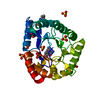
| |||||||||||||||
|---|---|---|---|---|---|---|---|---|---|---|---|---|---|---|---|---|
| 1 | 
| |||||||||||||||
| Unit cell |
| |||||||||||||||
| Components on special symmetry positions |
|
-Components
| #1: Protein | Mass: 29770.203 Da / Num. of mol.: 1 Source method: isolated from a genetically manipulated source Source: (gene. exp.) Clostridioides difficile (bacteria) / Gene: folP, SAMEA3374989_02220 / Production host: | ||||||||
|---|---|---|---|---|---|---|---|---|---|
| #2: Chemical | ChemComp-SO4 /   Mass: 96.063 Da / Num. of mol.: 9 / Source method: obtained synthetically / Formula: SO4 Mass: 96.063 Da / Num. of mol.: 9 / Source method: obtained synthetically / Formula: SO4#3: Chemical | ChemComp-PT1 / |   Mass: 312.283 Da / Num. of mol.: 1 / Source method: obtained synthetically / Formula: C14H12N6O3 / Feature type: SUBJECT OF INVESTIGATION Mass: 312.283 Da / Num. of mol.: 1 / Source method: obtained synthetically / Formula: C14H12N6O3 / Feature type: SUBJECT OF INVESTIGATION#4: Chemical | ChemComp-NHE / |   Mass: 207.290 Da / Num. of mol.: 1 / Source method: obtained synthetically / Formula: C8H17NO3S / Comment: pH buffer*YM Mass: 207.290 Da / Num. of mol.: 1 / Source method: obtained synthetically / Formula: C8H17NO3S / Comment: pH buffer*YM#5: Water | ChemComp-HOH / |  Mass: 18.015 Da / Num. of mol.: 148 / Source method: isolated from a natural source / Formula: H2O Mass: 18.015 Da / Num. of mol.: 148 / Source method: isolated from a natural source / Formula: H2OHas ligand of interest | Y | |
-Experimental details
-Experiment
| Experiment | Method: X-RAY DIFFRACTION / Number of used crystals: 1 |
|---|
- Sample preparation
| Crystal | Density Matthews: 5.01 Å3/Da / Density % sol: 75.47 % |
|---|---|
| Crystal grow | Temperature: 293 K / Method: vapor diffusion, hanging drop / pH: 9 Details: 2.4-2.6 M ammonium sulfate, 200 mM LiCl, 100 mM CHES (pH 9), 4 mM pteroic acid |
-Data collection
| Diffraction | Mean temperature: 100 K / Serial crystal experiment: N |
|---|---|
| Diffraction source | Source: SYNCHROTRON / Site: APS / Beamline: 19-BM / Wavelength: 0.99861 Å |
| Detector | Type: ADSC QUANTUM 210r / Detector: CCD / Date: Jun 6, 2018 |
| Radiation | Protocol: SINGLE WAVELENGTH / Monochromatic (M) / Laue (L): M / Scattering type: x-ray |
| Radiation wavelength | Wavelength: 0.99861 Å / Relative weight: 1 |
| Reflection | Resolution: 2→50 Å / Num. obs: 41365 / % possible obs: 99.2 % / Observed criterion σ(F): 0 / Observed criterion σ(I): 0 / Redundancy: 13 % / Rmerge(I) obs: 0.098 / Net I/σ(I): 45.4 |
| Reflection shell | Resolution: 2→2.03 Å / Redundancy: 7.7 % / Rmerge(I) obs: 0.454 / Mean I/σ(I) obs: 4.3 / Num. unique obs: 2026 / % possible all: 99.5 |
- Processing
| Software |
| ||||||||||||||||||||||||||||||||||||||||||||||||||||||||||||
|---|---|---|---|---|---|---|---|---|---|---|---|---|---|---|---|---|---|---|---|---|---|---|---|---|---|---|---|---|---|---|---|---|---|---|---|---|---|---|---|---|---|---|---|---|---|---|---|---|---|---|---|---|---|---|---|---|---|---|---|---|---|
| Refinement | Method to determine structure: MOLECULAR REPLACEMENT Starting model: 4hb7 Resolution: 2→37.56 Å / Cor.coef. Fo:Fc: 0.968 / Cor.coef. Fo:Fc free: 0.951 / SU B: 4.064 / SU ML: 0.1 / Cross valid method: THROUGHOUT / σ(F): 0 / ESU R: 0.106 / ESU R Free: 0.111 Details: HYDROGENS HAVE BEEN ADDED IN THE RIDING POSITIONS U VALUES : REFINED INDIVIDUALLY
| ||||||||||||||||||||||||||||||||||||||||||||||||||||||||||||
| Solvent computation | Ion probe radii: 0.8 Å / Shrinkage radii: 0.8 Å / VDW probe radii: 1.2 Å | ||||||||||||||||||||||||||||||||||||||||||||||||||||||||||||
| Displacement parameters | Biso max: 135.23 Å2 / Biso mean: 39.817 Å2 / Biso min: 23.66 Å2
| ||||||||||||||||||||||||||||||||||||||||||||||||||||||||||||
| Refinement step | Cycle: final / Resolution: 2→37.56 Å
| ||||||||||||||||||||||||||||||||||||||||||||||||||||||||||||
| Refine LS restraints |
| ||||||||||||||||||||||||||||||||||||||||||||||||||||||||||||
| LS refinement shell | Resolution: 2→2.048 Å / Rfactor Rfree error: 0
|
