 Movie
Movie Controller


+ Open data

- Basic information
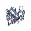
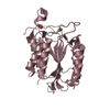
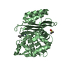
| Entry | Database: PDB / ID: 5fap | |||||||||||||||
|---|---|---|---|---|---|---|---|---|---|---|---|---|---|---|---|---|
| Title | CTX-M-15 in complex with FPI-1602 | |||||||||||||||
 Components Components | Beta-lactamase | |||||||||||||||
 Keywords Keywords | HYDROLASE/HYDROLASE INHIBITOR / HYDROLASE-HYDROLASE INHIBITOR complex | |||||||||||||||
| Function / homology |  Function and homology information Function and homology informationbeta-lactam antibiotic catabolic process / beta-lactamase activity / beta-lactamase / response to antibiotic Similarity search - Function | |||||||||||||||
| Biological species |  | |||||||||||||||
| Method |  X-RAY DIFFRACTION / SYNCHROTRON / MOLECULAR REPLACEMENT / Resolution: 2.7 Å X-RAY DIFFRACTION / SYNCHROTRON / MOLECULAR REPLACEMENT / Resolution: 2.7 Å | |||||||||||||||
 Authors Authors | King, A.M. / King, D.T. / French, S. / Brouillette, E. / Asli, A. / Alexander, A.N. / Vuckovic, M. / Maiti, S.N. / Parr, T.R. / Brown, E.D. ...King, A.M. / King, D.T. / French, S. / Brouillette, E. / Asli, A. / Alexander, A.N. / Vuckovic, M. / Maiti, S.N. / Parr, T.R. / Brown, E.D. / Malouin, F. / Strynadka, N.C.J. / Wright, G.D. | |||||||||||||||
| Funding support |  Canada, Canada,  United States, 4items United States, 4items
| |||||||||||||||
 Citation Citation | Journal: Acs Chem.Biol. / Year: 2016 Title: Structural and Kinetic Characterization of Diazabicyclooctanes as Dual Inhibitors of Both Serine-beta-Lactamases and Penicillin-Binding Proteins. Authors: King, A.M. / King, D.T. / French, S. / Brouillette, E. / Asli, A. / Alexander, J.A. / Vuckovic, M. / Maiti, S.N. / Parr, T.R. / Brown, E.D. / Malouin, F. / Strynadka, N.C. / Wright, G.D. | |||||||||||||||
| History |
|
- Structure visualization
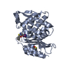
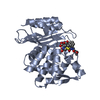
| Structure viewer | Molecule: MolmilJmol/JSmol |
|---|
- Downloads & links
-Download
| PDBx/mmCIF format | 5fap.cif.gz | 110.5 KB | Display | PDBx/mmCIF format |
|---|---|---|---|---|
| PDB format | pdb5fap.ent.gz | 84.9 KB | Display | PDB format |
| PDBx/mmJSON format | 5fap.json.gz | Tree view | PDBx/mmJSON format | |
| Others |  Other downloads Other downloads |
-Validation report
| Arichive directory | https://data.pdbj.org/pub/pdb/validation_reports/fa/5fapftp://data.pdbj.org/pub/pdb/validation_reports/fa/5fap | HTTPS FTP |
|---|
-Related structure data
| Related structure data |  5fa7C 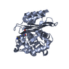 5faoC 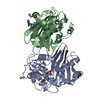 5faqC 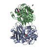 5fasC  5fatC  5fgzC 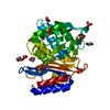 4hbtS C: citing same article ( S: Starting model for refinement |
|---|---|
| Similar structure data |


-Links
 PDBj
PDBj


- Assembly
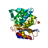
| Deposited unit | 
| ||||||||
|---|---|---|---|---|---|---|---|---|---|
| 1 | 
| ||||||||
| 2 | 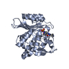
| ||||||||
| Unit cell |
|
-Components
| #1: Protein | Mass: 28138.826 Da / Num. of mol.: 2 Source method: isolated from a genetically manipulated source Source: (gene. exp.) Gene: blaUOE-1, bla, bla CTX-M-15, bla_1, bla_2, bla_3, blaCTX-M-15, blaCTX-M15, CTX-M-15, ECONIH1_02760, ECONIH1_27135, ERS085368_04262, ERS085370_01802, ERS085377_05268, ERS139214_01914, ERS139238_ ...Gene: blaUOE-1, bla, bla CTX-M-15, bla_1, bla_2, bla_3, blaCTX-M-15, blaCTX-M15, CTX-M-15, ECONIH1_02760, ECONIH1_27135, ERS085368_04262, ERS085370_01802, ERS085377_05268, ERS139214_01914, ERS139238_04648, ERS139238_04652, ERS150880_04508, HUS2011_pI0012, pCTXM15_EC8_00003, SK84_05077, SK86_03319 Production host: #2: Chemical |   Mass: 365.363 Da / Num. of mol.: 2 / Source method: obtained synthetically / Formula: C11H19N5O7S Mass: 365.363 Da / Num. of mol.: 2 / Source method: obtained synthetically / Formula: C11H19N5O7S#3: Water | ChemComp-HOH / |  Mass: 18.015 Da / Num. of mol.: 85 / Source method: isolated from a natural source / Formula: H2O Mass: 18.015 Da / Num. of mol.: 85 / Source method: isolated from a natural source / Formula: H2OHas protein modification | Y | |
|---|
-Experimental details
-Experiment
| Experiment | Method: X-RAY DIFFRACTION |
|---|
- Sample preparation
| Crystal | Density Matthews: 2.5 Å3/Da / Density % sol: 50.81 % |
|---|---|
| Crystal grow | Temperature: 298 K / Method: vapor diffusion, sitting drop / pH: 6.5 Details: 0.2M ammonium sulfate, 0.1M MES pH6.5, 30% PEG5K monomethyl ether |
-Data collection
| Diffraction | Mean temperature: 100 K |
|---|---|
| Diffraction source | Source: SYNCHROTRON / Site: CLSI / Beamline: 08ID-1 / Wavelength: 1 Å |
| Detector | Type: RAYONIX MX-300 / Detector: CCD / Date: Jul 15, 2014 |
| Radiation | Protocol: SINGLE WAVELENGTH / Monochromatic (M) / Laue (L): M / Scattering type: x-ray |
| Radiation wavelength | Wavelength: 1 Å / Relative weight: 1 |
| Reflection | Resolution: 2.7→30.97 Å / Num. obs: 14478 / % possible obs: 95.4 % / Redundancy: 2.5 % / Net I/σ(I): 11.7 |
- Processing
| Software |
| ||||||||||||||||||||||||||||||||||||||||||||||||||||||||||||||||||||||||||||||||||||||||||||||||||||||||||||||||||||||||||||||||||||||||||||||||||||||||||||||||||||||||||||||||||||||
|---|---|---|---|---|---|---|---|---|---|---|---|---|---|---|---|---|---|---|---|---|---|---|---|---|---|---|---|---|---|---|---|---|---|---|---|---|---|---|---|---|---|---|---|---|---|---|---|---|---|---|---|---|---|---|---|---|---|---|---|---|---|---|---|---|---|---|---|---|---|---|---|---|---|---|---|---|---|---|---|---|---|---|---|---|---|---|---|---|---|---|---|---|---|---|---|---|---|---|---|---|---|---|---|---|---|---|---|---|---|---|---|---|---|---|---|---|---|---|---|---|---|---|---|---|---|---|---|---|---|---|---|---|---|---|---|---|---|---|---|---|---|---|---|---|---|---|---|---|---|---|---|---|---|---|---|---|---|---|---|---|---|---|---|---|---|---|---|---|---|---|---|---|---|---|---|---|---|---|---|---|---|---|---|
| Refinement | Method to determine structure: MOLECULAR REPLACEMENT Starting model: 4HBT Resolution: 2.7→30.97 Å / Cor.coef. Fo:Fc: 0.93 / Cor.coef. Fo:Fc free: 0.889 / Cross valid method: THROUGHOUT / ESU R Free: 0.343 / Stereochemistry target values: MAXIMUM LIKELIHOOD / Details: HYDROGENS HAVE BEEN ADDED IN THE RIDING POSITIONS
| ||||||||||||||||||||||||||||||||||||||||||||||||||||||||||||||||||||||||||||||||||||||||||||||||||||||||||||||||||||||||||||||||||||||||||||||||||||||||||||||||||||||||||||||||||||||
| Solvent computation | Ion probe radii: 0.8 Å / Shrinkage radii: 0.8 Å / VDW probe radii: 1.2 Å / Solvent model: MASK | ||||||||||||||||||||||||||||||||||||||||||||||||||||||||||||||||||||||||||||||||||||||||||||||||||||||||||||||||||||||||||||||||||||||||||||||||||||||||||||||||||||||||||||||||||||||
| Displacement parameters | Biso mean: 27.48 Å2
| ||||||||||||||||||||||||||||||||||||||||||||||||||||||||||||||||||||||||||||||||||||||||||||||||||||||||||||||||||||||||||||||||||||||||||||||||||||||||||||||||||||||||||||||||||||||
| Refinement step | Cycle: LAST / Resolution: 2.7→30.97 Å
| ||||||||||||||||||||||||||||||||||||||||||||||||||||||||||||||||||||||||||||||||||||||||||||||||||||||||||||||||||||||||||||||||||||||||||||||||||||||||||||||||||||||||||||||||||||||
| Refine LS restraints |
|