 Movie
Movie Controller

[English] 日本語
 Yorodumi
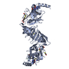
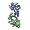
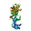
- PDB-1oph: NON-COVALENT COMPLEX BETWEEN ALPHA-1-PI-PITTSBURGH AND S195A TRYPSIN -
+ Open data

- Basic information
| Entry | Database: PDB / ID: 1oph | ||||||
|---|---|---|---|---|---|---|---|
| Title | NON-COVALENT COMPLEX BETWEEN ALPHA-1-PI-PITTSBURGH AND S195A TRYPSIN | ||||||
 Components Components |
| ||||||
 Keywords Keywords | HYDROLASE/HYDROLASE INHIBITOR / SERINE PROTEINASE INHIBITOR / MICHAELIS-LIKE COMPLEX / SERPIN / ALPHA-1 ANTITRYPSIN / TRYPSIN / HYDROLASE-HYDROLASE INHIBITOR COMPLEX | ||||||
| Function / homology |  Function and homology information Function and homology informationCargo concentration in the ER / COPII-coated ER to Golgi transport vesicle / COPII-mediated vesicle transport / trypsin / serpin family protein binding / serine protease inhibitor complex / digestion / endoplasmic reticulum-Golgi intermediate compartment membrane / platelet alpha granule lumen / acute-phase response ...Cargo concentration in the ER / COPII-coated ER to Golgi transport vesicle / COPII-mediated vesicle transport / trypsin / serpin family protein binding / serine protease inhibitor complex / digestion / endoplasmic reticulum-Golgi intermediate compartment membrane / platelet alpha granule lumen / acute-phase response / Post-translational protein phosphorylation / serine-type endopeptidase inhibitor activity / Regulation of Insulin-like Growth Factor (IGF) transport and uptake by Insulin-like Growth Factor Binding Proteins (IGFBPs) / blood coagulation / Platelet degranulation / extracellular matrix / protease binding / endopeptidase activity / ficolin-1-rich granule lumen / endoplasmic reticulum lumen / serine-type endopeptidase activity / Neutrophil degranulation / Golgi apparatus / endoplasmic reticulum / proteolysis / : / extracellular exosome / extracellular region / metal ion binding / identical protein binding Similarity search - Function | ||||||
| Biological species |  Homo sapiens (human) Homo sapiens (human) | ||||||
| Method |  X-RAY DIFFRACTION / SYNCHROTRON / MOLECULAR REPLACEMENT / Resolution: 2.3 Å X-RAY DIFFRACTION / SYNCHROTRON / MOLECULAR REPLACEMENT / Resolution: 2.3 Å | ||||||
 Authors Authors | Dementiev, A. / Simonovic, M. / Volz, K. / Gettins, P.G. | ||||||
 Citation Citation | Journal: J.Biol.Chem. / Year: 2003 Title: Canonical inhibitor-like interactions explain reactivity of alpha1-proteinase inhibitor Pittsburgh and antithrombin with proteinases Authors: Dementiev, A. / Simonovic, M. / Volz, K. / Gettins, P.G. | ||||||
| History |
|
- Structure visualization
| Structure viewer | Molecule: MolmilJmol/JSmol |
|---|
- Downloads & links
-Download
| PDBx/mmCIF format | 1oph.cif.gz | 133.7 KB | Display | PDBx/mmCIF format |
|---|---|---|---|---|
| PDB format | pdb1oph.ent.gz | 103.1 KB | Display | PDB format |
| PDBx/mmJSON format | 1oph.json.gz | Tree view | PDBx/mmJSON format | |
| Others |  Other downloads Other downloads |
-Validation report
| Arichive directory | https://data.pdbj.org/pub/pdb/validation_reports/op/1ophftp://data.pdbj.org/pub/pdb/validation_reports/op/1oph | HTTPS FTP |
|---|
-Related structure data
| Related structure data | 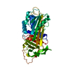 1oo8C  1ejmS  1qlpS C: citing same article ( S: Starting model for refinement |
|---|---|
| Similar structure data |


-Links
 PDBj
PDBj


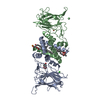
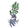
- Assembly
| Deposited unit | 
| ||||||||
|---|---|---|---|---|---|---|---|---|---|
| 1 |
| ||||||||
| 2 | 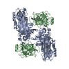
| ||||||||
| Unit cell |
|
-Components
| #1: Protein | Mass: 44276.258 Da / Num. of mol.: 1 / Fragment: alpha-1-antitrypsin (RESIDUES 26-418) Mutation: F51L, T59A, T68A, A70G, C232S, M358R, M274I, S381A, K387R Source method: isolated from a genetically manipulated source Source: (gene. exp.) Homo sapiens (human) / Tissue: BLOOD / Gene: SERPINA1 OR PI OR AAT / Plasmid: pQE30 / Cell line (production host): SG13009 / Production host:  |
|---|---|
| #2: Protein | Mass: 25428.717 Da / Num. of mol.: 1 / Mutation: S195A Source method: isolated from a genetically manipulated source Source: (gene. exp.) |
| #3: Water | ChemComp-HOH /  Mass: 18.015 Da / Num. of mol.: 377 / Source method: isolated from a natural source / Formula: H2O Mass: 18.015 Da / Num. of mol.: 377 / Source method: isolated from a natural source / Formula: H2O |
| Has protein modification | Y |
-Experimental details
-Experiment
| Experiment | Method: X-RAY DIFFRACTION / Number of used crystals: 2 |
|---|
- Sample preparation
| Crystal | Density Matthews: 2.77 Å3/Da / Density % sol: 55.31 % | ||||||||||||||||||||||||
|---|---|---|---|---|---|---|---|---|---|---|---|---|---|---|---|---|---|---|---|---|---|---|---|---|---|
| Crystal grow | Temperature: 291 K / Method: vapor diffusion, hanging drop / pH: 8.3 Details: 0.2 M tri-K-citrate, 17.5% PEG 3350, 2% GLYCEROL, pH 8.30, VAPOR DIFFUSION, HANGING DROP, temperature 291K | ||||||||||||||||||||||||
| Crystal grow | *PLUS Temperature: 18 ℃ / Method: vapor diffusion, hanging drop / PH range low: 8.5 / PH range high: 8.3 | ||||||||||||||||||||||||
| Components of the solutions | *PLUS
|
-Data collection
| Diffraction |
| ||||||||||||||||||
|---|---|---|---|---|---|---|---|---|---|---|---|---|---|---|---|---|---|---|---|
| Diffraction source |
| ||||||||||||||||||
| Detector |
| ||||||||||||||||||
| Radiation |
| ||||||||||||||||||
| Radiation wavelength | Wavelength: 1 Å / Relative weight: 1 | ||||||||||||||||||
| Reflection | Resolution: 2.3→40 Å / Num. all: 32771 / Num. obs: 32771 / % possible obs: 97.5 % / Observed criterion σ(F): 0 / Observed criterion σ(I): 0 / Redundancy: 19.2 % / Biso Wilson estimate: 27.4 Å2 / Rmerge(I) obs: 0.103 / Net I/σ(I): 20.6 | ||||||||||||||||||
| Reflection shell | Resolution: 2.3→2.38 Å / Rmerge(I) obs: 0.289 / Mean I/σ(I) obs: 3.75 / Num. unique all: 3330 / % possible all: 97.3 | ||||||||||||||||||
| Reflection | *PLUS Highest resolution: 2.3 Å / Lowest resolution: 100 Å / Num. obs: 33539 / % possible obs: 99.5 % / Num. measured all: 628540 / Rmerge(I) obs: 0.096 | ||||||||||||||||||
| Reflection shell | *PLUS % possible obs: 99.7 % / Rmerge(I) obs: 0.245 / Mean I/σ(I) obs: 4.1 |

- Processing
| Software |
| ||||||||||||||||||||||||||||||||||||
|---|---|---|---|---|---|---|---|---|---|---|---|---|---|---|---|---|---|---|---|---|---|---|---|---|---|---|---|---|---|---|---|---|---|---|---|---|---|
| Refinement | Method to determine structure: MOLECULAR REPLACEMENT Starting model: PDB ENTRY 1QLP AND 1EJM Resolution: 2.3→22.51 Å / Rfactor Rfree error: 0.004 / Isotropic thermal model: RESTRAINED / Cross valid method: THROUGHOUT / σ(F): 0 / Stereochemistry target values: Engh & Huber
| ||||||||||||||||||||||||||||||||||||
| Solvent computation | Solvent model: FLAT MODEL / Bsol: 49.0933 Å2 / ksol: 0.356808 e/Å3 | ||||||||||||||||||||||||||||||||||||
| Displacement parameters | Biso mean: 42 Å2
| ||||||||||||||||||||||||||||||||||||
| Refine analyze |
| ||||||||||||||||||||||||||||||||||||
| Refinement step | Cycle: LAST / Resolution: 2.3→22.51 Å
| ||||||||||||||||||||||||||||||||||||
| Refine LS restraints |
| ||||||||||||||||||||||||||||||||||||
| LS refinement shell | Resolution: 2.3→2.41 Å / Rfactor Rfree error: 0.013 / Total num. of bins used: 6
| ||||||||||||||||||||||||||||||||||||
| Xplor file |
| ||||||||||||||||||||||||||||||||||||
| Refinement | *PLUS Lowest resolution: 40 Å / Rfactor Rfree: 0.228 / Rfactor Rwork: 0.189 | ||||||||||||||||||||||||||||||||||||
| Solvent computation | *PLUS | ||||||||||||||||||||||||||||||||||||
| Displacement parameters | *PLUS | ||||||||||||||||||||||||||||||||||||
| Refine LS restraints | *PLUS
|
