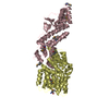
 Movie
Movie Controller


+ Open data

- Basic information
| Entry | Database: PDB / ID: 4ubf | ||||||
|---|---|---|---|---|---|---|---|
| Title | HsMCAK motor domain complex | ||||||
 Components Components | (Kinesin-like protein KIF2C) x 2 | ||||||
 Keywords Keywords | CELL CYCLE / MCAK / Kif2c / Complex / Motor domain | ||||||
| Function / homology |  Function and homology information Function and homology informationpostsynaptic cytoskeleton organization / regulation of chromosome segregation / establishment or maintenance of microtubule cytoskeleton polarity / metaphase chromosome alignment / centromeric DNA binding / microtubule plus-end / attachment of mitotic spindle microtubules to kinetochore / microtubule plus-end binding / Kinesins / microtubule depolymerization ...postsynaptic cytoskeleton organization / regulation of chromosome segregation / establishment or maintenance of microtubule cytoskeleton polarity / metaphase chromosome alignment / centromeric DNA binding / microtubule plus-end / attachment of mitotic spindle microtubules to kinetochore / microtubule plus-end binding / Kinesins / microtubule depolymerization / microtubule motor activity / kinesin complex / COPI-dependent Golgi-to-ER retrograde traffic / microtubule-based movement / regulation of neurotransmitter receptor localization to postsynaptic specialization membrane / mitotic metaphase chromosome alignment / chromosome, centromeric region / Amplification of signal from unattached kinetochores via a MAD2 inhibitory signal / MHC class II antigen presentation / Mitotic Prometaphase / EML4 and NUDC in mitotic spindle formation / Resolution of Sister Chromatid Cohesion / RHO GTPases Activate Formins / kinetochore / spindle / Separation of Sister Chromatids / microtubule cytoskeleton / presynapse / microtubule binding / microtubule / postsynapse / cell division / centrosome / glutamatergic synapse / ATP hydrolysis activity / ATP binding / membrane / nucleus / cytosol / cytoplasm Similarity search - Function | ||||||
| Biological species |  Homo sapiens (human) Homo sapiens (human) | ||||||
| Method |  X-RAY DIFFRACTION / SYNCHROTRON / MOLECULAR REPLACEMENT / Resolution: 3 Å X-RAY DIFFRACTION / SYNCHROTRON / MOLECULAR REPLACEMENT / Resolution: 3 Å | ||||||
 Authors Authors | Welburn, J.P.I. / Talapatra, S.K. | ||||||
| Funding support |  United Kingdom, 1items United Kingdom, 1items
| ||||||
 Citation Citation | Journal: Elife / Year: 2015 Title: The C-terminal region of the motor protein MCAK controls its structure and activity through a conformational switch. Authors: Talapatra, S.K. / Harker, B. / Welburn, J.P. | ||||||
| History |
|
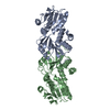
- Structure visualization
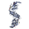
| Structure viewer | Molecule: MolmilJmol/JSmol |
|---|
- Downloads & links
-Download
| PDBx/mmCIF format | 4ubf.cif.gz | 264.5 KB | Display | PDBx/mmCIF format |
|---|---|---|---|---|
| PDB format | pdb4ubf.ent.gz | 206.1 KB | Display | PDB format |
| PDBx/mmJSON format | 4ubf.json.gz | Tree view | PDBx/mmJSON format | |
| Others |  Other downloads Other downloads |
-Validation report
| Arichive directory | https://data.pdbj.org/pub/pdb/validation_reports/ub/4ubfftp://data.pdbj.org/pub/pdb/validation_reports/ub/4ubf | HTTPS FTP |
|---|
-Related structure data
| Related structure data | 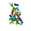 2hehS S: Starting model for refinement |
|---|---|
| Similar structure data |


-Links
 PDBj
PDBj


- Assembly
| Deposited unit | 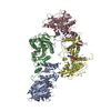
| ||||||||
|---|---|---|---|---|---|---|---|---|---|
| 1 | 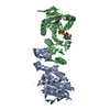
| ||||||||
| 2 | 
| ||||||||
| Unit cell |
|
-Components
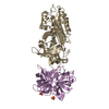
| #1: Protein | Mass: 43295.602 Da / Num. of mol.: 4 / Fragment: UNP Residues 225-593 Source method: isolated from a genetically manipulated source Source: (gene. exp.) Homo sapiens (human) / Gene: KIF2C, KNSL6 / Details (production host): pET28a-LIC / Production host:  #2: Protein/peptide | | Mass: 1376.450 Da / Num. of mol.: 1 / Fragment: UNP Residues 709-720 Source method: isolated from a genetically manipulated source Source: (gene. exp.) Homo sapiens (human) / Gene: KIF2C, KNSL6 / Plasmid: pET28a-LIC / Production host: #3: Chemical | ChemComp-MG /   Mass: 24.305 Da / Num. of mol.: 7 / Source method: isolated from a natural source / Formula: Mg / Source: (natural) Homo sapiens (human) Mass: 24.305 Da / Num. of mol.: 7 / Source method: isolated from a natural source / Formula: Mg / Source: (natural) Homo sapiens (human)#4: Chemical | ChemComp-ADP /   Mass: 427.201 Da / Num. of mol.: 4 / Source method: obtained synthetically / Formula: C10H15N5O10P2 / Comment: ADP, energy-carrying molecule*YM Mass: 427.201 Da / Num. of mol.: 4 / Source method: obtained synthetically / Formula: C10H15N5O10P2 / Comment: ADP, energy-carrying molecule*YM#5: Water | ChemComp-HOH / |  Mass: 18.015 Da / Num. of mol.: 69 / Source method: isolated from a natural source / Formula: H2O Mass: 18.015 Da / Num. of mol.: 69 / Source method: isolated from a natural source / Formula: H2O |
|---|
-Experimental details
-Experiment
| Experiment | Method: X-RAY DIFFRACTION |
|---|
- Sample preparation
| Crystal | Density Matthews: 2.61 Å3/Da / Density % sol: 52.94 % / Description: Single Rectangular Crystal |
|---|---|
| Crystal grow | Temperature: 292 K / Method: vapor diffusion, sitting drop / pH: 6.8 Details: 24 % w/v PEG 1500 20 % v/v Glycerol Protein crystals appeared after two days and single crystals were used for measurement Temp details: Controlled Temperature Room |
-Data collection
| Diffraction | Mean temperature: 80 K |
|---|---|
| Diffraction source | Source: SYNCHROTRON / Site: Diamond / Beamline: I24 / Wavelength: 0.987 Å |
| Detector | Type: PSI PILATUS 6M / Detector: PIXEL / Date: Jul 11, 2014 |
| Radiation | Protocol: SINGLE WAVELENGTH / Monochromatic (M) / Laue (L): M / Scattering type: x-ray |
| Radiation wavelength | Wavelength: 0.987 Å / Relative weight: 1 |
| Reflection | Resolution: 3→30 Å / Num. all: 155983 / Num. obs: 35146 / % possible obs: 99.8 % / Redundancy: 4.4 % / Rmerge(I) obs: 0.091 / Net I/σ(I): 10.2 |
| Reflection shell | Resolution: 3→3.5 Å / Redundancy: 4.5 % / Rmerge(I) obs: 0.682 / Mean I/σ(I) obs: 2 / % possible all: 100 |
- Processing
| Software | Name: PHENIX / Version: (phenix.refine: 1.8.2_1309) / Classification: refinement | |||||||||||||||||||||||||||||||||||||||||||||||||||||||||||||||||||||||||||||||||||||||||||
|---|---|---|---|---|---|---|---|---|---|---|---|---|---|---|---|---|---|---|---|---|---|---|---|---|---|---|---|---|---|---|---|---|---|---|---|---|---|---|---|---|---|---|---|---|---|---|---|---|---|---|---|---|---|---|---|---|---|---|---|---|---|---|---|---|---|---|---|---|---|---|---|---|---|---|---|---|---|---|---|---|---|---|---|---|---|---|---|---|---|---|---|---|
| Refinement | Method to determine structure: MOLECULAR REPLACEMENT Starting model: 2HEH Resolution: 3→29.492 Å / SU ML: 0.52 / Cross valid method: FREE R-VALUE / σ(F): 1.34 / Phase error: 33.46 / Stereochemistry target values: ML
| |||||||||||||||||||||||||||||||||||||||||||||||||||||||||||||||||||||||||||||||||||||||||||
| Solvent computation | Shrinkage radii: 0.9 Å / VDW probe radii: 1.11 Å / Solvent model: FLAT BULK SOLVENT MODEL | |||||||||||||||||||||||||||||||||||||||||||||||||||||||||||||||||||||||||||||||||||||||||||
| Refinement step | Cycle: LAST / Resolution: 3→29.492 Å
| |||||||||||||||||||||||||||||||||||||||||||||||||||||||||||||||||||||||||||||||||||||||||||
| Refine LS restraints |
| |||||||||||||||||||||||||||||||||||||||||||||||||||||||||||||||||||||||||||||||||||||||||||
| LS refinement shell |
|
