 Movie
Movie Controller

[English] 日本語
 Yorodumi
- PDB-3pb1: Crystal Structure of a Michaelis Complex between Plasminogen Acti... -
+ Open data

- Basic information
| Entry | Database: PDB / ID: 3pb1 | ||||||
|---|---|---|---|---|---|---|---|
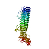
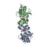
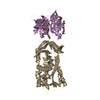
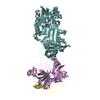
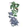
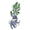
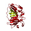
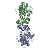
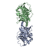
| Title | Crystal Structure of a Michaelis Complex between Plasminogen Activator Inhibitor-1 and Urokinase-type Plasminogen Activator | ||||||
 Components Components |
| ||||||
 Keywords Keywords | hydrolase Inhibitor/Hydrolase / PAI-1 / uPA / Michaelis complex / Structural Genomics / Structure 2 Function Project / S2F / hydrolase Inhibitor-Hydrolase complex | ||||||
| Function / homology |  Function and homology information Function and homology informationpositive regulation of leukotriene production involved in inflammatory response / negative regulation of smooth muscle cell-matrix adhesion / negative regulation of integrin-mediated signaling pathway / peptidase inhibitor complex / dentinogenesis / positive regulation of coagulation / negative regulation of vascular wound healing / negative regulation of smooth muscle cell migration / Regulation of MITF-M-dependent genes involved in extracellular matrix, focal adhesion and epithelial-to-mesenchymal transition / negative regulation of wound healing ...positive regulation of leukotriene production involved in inflammatory response / negative regulation of smooth muscle cell-matrix adhesion / negative regulation of integrin-mediated signaling pathway / peptidase inhibitor complex / dentinogenesis / positive regulation of coagulation / negative regulation of vascular wound healing / negative regulation of smooth muscle cell migration / Regulation of MITF-M-dependent genes involved in extracellular matrix, focal adhesion and epithelial-to-mesenchymal transition / negative regulation of wound healing / positive regulation of odontoblast differentiation / u-plasminogen activator / regulation of smooth muscle cell-matrix adhesion / urokinase plasminogen activator signaling pathway / regulation of plasminogen activation / regulation of integrin-mediated signaling pathway / protein complex involved in cell-matrix adhesion / regulation of fibrinolysis / regulation of wound healing / negative regulation of plasminogen activation / serine-type endopeptidase complex / regulation of smooth muscle cell migration / Dissolution of Fibrin Clot / negative regulation of cell adhesion mediated by integrin / regulation of cell adhesion mediated by integrin / positive regulation of monocyte chemotaxis / smooth muscle cell migration / plasminogen activation / endopeptidase inhibitor activity / negative regulation of thrombin-activated receptor signaling pathway / tertiary granule membrane / replicative senescence / negative regulation of blood coagulation / negative regulation of fibrinolysis / positive regulation of blood coagulation / negative regulation of endothelial cell apoptotic process / ECM proteoglycans / regulation of cell adhesion / negative regulation of extrinsic apoptotic signaling pathway via death domain receptors / serine protease inhibitor complex / specific granule membrane / fibrinolysis / positive regulation of epidermal growth factor receptor signaling pathway / negative regulation of proteolysis / BMAL1:CLOCK,NPAS2 activates circadian expression / platelet alpha granule lumen / negative regulation of cell migration / positive regulation of interleukin-8 production / serine-type endopeptidase inhibitor activity / SMAD2/SMAD3:SMAD4 heterotrimer regulates transcription / positive regulation of receptor-mediated endocytosis / chemotaxis / positive regulation of angiogenesis / blood coagulation / positive regulation of inflammatory response / Platelet degranulation / regulation of cell population proliferation / cellular response to lipopolysaccharide / extracellular matrix / protease binding / angiogenesis / defense response to Gram-negative bacterium / response to hypoxia / positive regulation of cell migration / receptor ligand activity / serine-type endopeptidase activity / external side of plasma membrane / signaling receptor binding / focal adhesion / Neutrophil degranulation / cell surface / signal transduction / proteolysis / : / extracellular exosome / extracellular region / plasma membrane Similarity search - Function | ||||||
| Biological species |  Homo sapiens (human) Homo sapiens (human) | ||||||
| Method |  X-RAY DIFFRACTION / SYNCHROTRON / MOLECULAR REPLACEMENT / Resolution: 2.3 Å X-RAY DIFFRACTION / SYNCHROTRON / MOLECULAR REPLACEMENT / Resolution: 2.3 Å | ||||||
 Authors Authors | Lin, Z. / Jiang, L. / Huang, M. / Structure 2 Function Project (S2F) | ||||||
 Citation Citation | Journal: J.Biol.Chem. / Year: 2011 Title: Structural basis for recognition of urokinase-type plasminogen activator by plasminogen activator inhibitor-1. Authors: Lin, Z. / Jiang, L. / Yuan, C. / Jensen, J.K. / Zhang, X. / Luo, Z. / Furie, B.C. / Furie, B. / Andreasen, P.A. / Huang, M. | ||||||
| History |
|
- Structure visualization
| Structure viewer | Molecule: MolmilJmol/JSmol |
|---|
- Downloads & links
-Download
| PDBx/mmCIF format | 3pb1.cif.gz | 141.7 KB | Display | PDBx/mmCIF format |
|---|---|---|---|---|
| PDB format | pdb3pb1.ent.gz | 108.9 KB | Display | PDB format |
| PDBx/mmJSON format | 3pb1.json.gz | Tree view | PDBx/mmJSON format | |
| Others |  Other downloads Other downloads |
-Validation report
| Arichive directory | https://data.pdbj.org/pub/pdb/validation_reports/pb/3pb1ftp://data.pdbj.org/pub/pdb/validation_reports/pb/3pb1 | HTTPS FTP |
|---|
-Related structure data
| Related structure data |  1dvmS S: Starting model for refinement |
|---|---|
| Similar structure data |


-Links
 PDBj
PDBj


- Assembly
| Deposited unit | 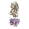
| ||||||||
|---|---|---|---|---|---|---|---|---|---|
| 1 |
| ||||||||
| Unit cell |
|
-Components
| #1: Protein | Mass: 42795.066 Da / Num. of mol.: 1 / Mutation: N150H, K154T, Q319L, M354I Source method: isolated from a genetically manipulated source Source: (gene. exp.) Homo sapiens (human) / Gene: SERPINE1, PAI1, PLANH1 / Production host:  | ||||
|---|---|---|---|---|---|
| #2: Protein | Mass: 28426.373 Da / Num. of mol.: 1 / Fragment: UNP RESIDUES 143-395 / Mutation: S195A Source method: isolated from a genetically manipulated source Source: (gene. exp.) Homo sapiens (human) / Gene: PLAU, RP11-417O11.1-002 / Production host:  Pichia pastoris (fungus) Pichia pastoris (fungus)References: UniProt: Q5SWW8, UniProt: P00749*PLUS, u-plasminogen activator | ||||
| #3: Chemical |   Mass: 96.063 Da / Num. of mol.: 2 / Source method: obtained synthetically / Formula: SO4 Mass: 96.063 Da / Num. of mol.: 2 / Source method: obtained synthetically / Formula: SO4#4: Water | ChemComp-HOH / |  Mass: 18.015 Da / Num. of mol.: 314 / Source method: isolated from a natural source / Formula: H2O Mass: 18.015 Da / Num. of mol.: 314 / Source method: isolated from a natural source / Formula: H2OHas protein modification | Y | |
-Experimental details
-Experiment
| Experiment | Method: X-RAY DIFFRACTION / Number of used crystals: 1 |
|---|
- Sample preparation
| Crystal | Density Matthews: 3.33 Å3/Da / Density % sol: 63.02 % |
|---|---|
| Crystal grow | Temperature: 293 K / Method: vapor diffusion, hanging drop / pH: 7.4 Details: 1.4M ammonium sulfate, 0.1M Tris-HCl, pH 7.4, VAPOR DIFFUSION, HANGING DROP, temperature 293K |
-Data collection
| Diffraction | Mean temperature: 100 K |
|---|---|
| Diffraction source | Source: SYNCHROTRON / Site: SSRF  / Beamline: BL17U / Wavelength: 1 Å / Beamline: BL17U / Wavelength: 1 Å |
| Detector | Detector: DIFFRACTOMETER / Date: Jun 16, 2010 |
| Radiation | Protocol: SINGLE WAVELENGTH / Monochromatic (M) / Laue (L): M / Scattering type: x-ray |
| Radiation wavelength | Wavelength: 1 Å / Relative weight: 1 |
| Reflection | Resolution: 2.3→99 Å / Num. all: 81438 / Num. obs: 80462 / % possible obs: 99.9 % / Redundancy: 5.9 % / Rmerge(I) obs: 0.162 / Rsym value: 0.185 / Net I/σ(I): 21.5 |
| Reflection shell | Resolution: 2.3→2.34 Å / Redundancy: 4.2 % / Rmerge(I) obs: 0.589 / Mean I/σ(I) obs: 3.4 / Num. unique all: 3996 / Rsym value: 0.633 / % possible all: 98.5 |
- Processing
| Software |
| |||||||||||||||||||||||||||||||||||||||||||||||||||||||||||||||||
|---|---|---|---|---|---|---|---|---|---|---|---|---|---|---|---|---|---|---|---|---|---|---|---|---|---|---|---|---|---|---|---|---|---|---|---|---|---|---|---|---|---|---|---|---|---|---|---|---|---|---|---|---|---|---|---|---|---|---|---|---|---|---|---|---|---|---|
| Refinement | Method to determine structure: MOLECULAR REPLACEMENT Starting model: PDB ENTRY 1DVM Resolution: 2.3→38.32 Å / Cor.coef. Fo:Fc: 0.925 / Cor.coef. Fo:Fc free: 0.891 / SU B: 6.525 / SU ML: 0.16 / Cross valid method: THROUGHOUT / ESU R Free: 0.23 / Stereochemistry target values: MAXIMUM LIKELIHOOD / Details: HYDROGENS HAVE BEEN ADDED IN THE RIDING POSITIONS
| |||||||||||||||||||||||||||||||||||||||||||||||||||||||||||||||||
| Solvent computation | Ion probe radii: 0.8 Å / Shrinkage radii: 0.8 Å / VDW probe radii: 1.4 Å / Solvent model: MASK | |||||||||||||||||||||||||||||||||||||||||||||||||||||||||||||||||
| Displacement parameters | Biso mean: 28.466 Å2
| |||||||||||||||||||||||||||||||||||||||||||||||||||||||||||||||||
| Refinement step | Cycle: LAST / Resolution: 2.3→38.32 Å
| |||||||||||||||||||||||||||||||||||||||||||||||||||||||||||||||||
| Refine LS restraints |
| |||||||||||||||||||||||||||||||||||||||||||||||||||||||||||||||||
| LS refinement shell | Resolution: 2.301→2.361 Å / Total num. of bins used: 20
|
