 Movie
Movie Controller

[English] 日本語
 Yorodumi
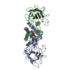
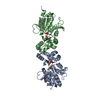
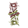
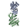
- PDB-3zwk: The 3-dimensional structure of MpgP from Thermus thermophilus HB2... -
+ Open data

- Basic information
| Entry | Database: PDB / ID: 3zwk | ||||||
|---|---|---|---|---|---|---|---|
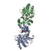
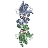
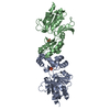
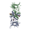
| Title | The 3-dimensional structure of MpgP from Thermus thermophilus HB27, in complex with the metavanadate | ||||||
 Components Components | MANNOSYL-3-PHOSPHOGLYCERATE PHOSPHATASE | ||||||
 Keywords Keywords | HYDROLASE / HALOALKANOID ACID DEHALOGENASE-LIKE PHOSPHATASE / HAD-LIKE PHOSPHATASE / CRYSTALLOGRAPHIC SNAPSHOT | ||||||
| Function / homology |  Function and homology information Function and homology informationmannosyl-3-phosphoglycerate phosphatase / mannosyl-3-phosphoglycerate phosphatase activity / mannosylglycerate biosynthetic process / magnesium ion binding / cytosol Similarity search - Function | ||||||
| Biological species |   THERMUS THERMOPHILUS (bacteria) THERMUS THERMOPHILUS (bacteria) | ||||||
| Method |  X-RAY DIFFRACTION / SYNCHROTRON / MOLECULAR REPLACEMENT / Resolution: 2.099 Å X-RAY DIFFRACTION / SYNCHROTRON / MOLECULAR REPLACEMENT / Resolution: 2.099 Å | ||||||
 Authors Authors | Goncalves, S. / Esteves, A.M. / Santos, H. / Borges, N. / Matias, P.M. | ||||||
 Citation Citation | Journal: Biochemistry / Year: 2011 Title: The Three-Dimensional Structure of Mannosyl-3-Phosphoglycerate Phosphatase from Thermus Thermophilus Hb27: A New Member of the Haloalkanoic Acid Dehalogenase Superfamily. Authors: Goncalves, S. / Esteves, A.M. / Santos, H. / Borges, N. / Matias, P.M. #1: Journal: Acta Crystallogr.,Sect.F / Year: 2011 Title: Crystallization and Preliminary X-Ray Analysis of Mannosyl-3-Phosphoglycerate Phosphatase from Thermus Thermophilus Hb27. Authors: Goncalves, S. / Esteves, A.M. / Borges, N. / Santos, H. / Matias, P.M. | ||||||
| History |
|
- Structure visualization
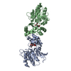
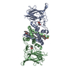
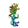
| Structure viewer | Molecule: MolmilJmol/JSmol |
|---|
- Downloads & links
-Download
| PDBx/mmCIF format | 3zwk.cif.gz | 215 KB | Display | PDBx/mmCIF format |
|---|---|---|---|---|
| PDB format | pdb3zwk.ent.gz | 174.7 KB | Display | PDB format |
| PDBx/mmJSON format | 3zwk.json.gz | Tree view | PDBx/mmJSON format | |
| Others |  Other downloads Other downloads |
-Validation report
| Arichive directory | https://data.pdbj.org/pub/pdb/validation_reports/zw/3zwkftp://data.pdbj.org/pub/pdb/validation_reports/zw/3zwk | HTTPS FTP |
|---|
-Related structure data
| Related structure data |  3ztwC 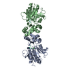 3ztySC  3zu6C  3zupC  3zw7C  3zwdC  3zx4C  3zx5C C: citing same article ( S: Starting model for refinement |
|---|---|
| Similar structure data |

-Links
 PDBj
PDBj

- Assembly
| Deposited unit | 
| ||||||||
|---|---|---|---|---|---|---|---|---|---|
| 1 |
| ||||||||
| Unit cell |
| ||||||||
| Noncrystallographic symmetry (NCS) | NCS oper: (Code: given Matrix: (0.80856, 0.00988, 0.58833), Vector: |
-Components
| #1: Protein | Mass: 28219.281 Da / Num. of mol.: 2 Source method: isolated from a genetically manipulated source Source: (gene. exp.) THERMUS THERMOPHILUS (bacteria) / Strain: HB27 / Plasmid: PKK223-3 / Production host: References: UniProt: Q72K29, mannosyl-3-phosphoglycerate phosphatase #2: Chemical |   Mass: 24.305 Da / Num. of mol.: 2 / Source method: obtained synthetically / Formula: Mg Mass: 24.305 Da / Num. of mol.: 2 / Source method: obtained synthetically / Formula: Mg#3: Chemical | ChemComp-VN3 / |   Mass: 98.940 Da / Num. of mol.: 1 / Source method: obtained synthetically / Formula: VO3 Mass: 98.940 Da / Num. of mol.: 1 / Source method: obtained synthetically / Formula: VO3#4: Water | ChemComp-HOH / |  Mass: 18.015 Da / Num. of mol.: 427 / Source method: isolated from a natural source / Formula: H2O Mass: 18.015 Da / Num. of mol.: 427 / Source method: isolated from a natural source / Formula: H2OHas protein modification | Y | |
|---|
-Experimental details
-Experiment
| Experiment | Method: X-RAY DIFFRACTION / Number of used crystals: 1 |
|---|
- Sample preparation
| Crystal | Density Matthews: 2.42 Å3/Da / Density % sol: 49.12 % / Description: NONE |
|---|---|
| Crystal grow | pH: 6.5 / Details: pH 6.5 |
-Data collection
| Diffraction | Mean temperature: 100 K |
|---|---|
| Diffraction source | Source: SYNCHROTRON / Site: ESRF  / Beamline: ID23-1 / Wavelength: 0.97925 / Beamline: ID23-1 / Wavelength: 0.97925 |
| Detector | Type: ADSC CCD / Detector: CCD / Date: Sep 10, 2010 / Details: BENT CYLINDRICAL MIRROR |
| Radiation | Monochromator: SI (111) / Protocol: SINGLE WAVELENGTH / Monochromatic (M) / Laue (L): M / Scattering type: x-ray |
| Radiation wavelength | Wavelength: 0.97925 Å / Relative weight: 1 |
| Reflection | Resolution: 2.1→60 Å / Num. obs: 30243 / % possible obs: 99 % / Observed criterion σ(I): -3 / Redundancy: 3.4 % / Biso Wilson estimate: 30.62 Å2 / Rmerge(I) obs: 0.05 / Net I/σ(I): 16.78 |
| Reflection shell | Resolution: 2.1→2.23 Å / Redundancy: 3.1 % / Rmerge(I) obs: 0.29 / Mean I/σ(I) obs: 4.47 / % possible all: 96.5 |
- Processing
| Software |
| |||||||||||||||||||||||||||||||||||||||||||||||||||||||||||||||||||||||||||||||||||||||||||||||||||||||||||||||||||||||||||||||||||||||||||||||||||||||||||||||||||||||||||||||||||||||||||||||||||||||||||||||||||||||||||||||||
|---|---|---|---|---|---|---|---|---|---|---|---|---|---|---|---|---|---|---|---|---|---|---|---|---|---|---|---|---|---|---|---|---|---|---|---|---|---|---|---|---|---|---|---|---|---|---|---|---|---|---|---|---|---|---|---|---|---|---|---|---|---|---|---|---|---|---|---|---|---|---|---|---|---|---|---|---|---|---|---|---|---|---|---|---|---|---|---|---|---|---|---|---|---|---|---|---|---|---|---|---|---|---|---|---|---|---|---|---|---|---|---|---|---|---|---|---|---|---|---|---|---|---|---|---|---|---|---|---|---|---|---|---|---|---|---|---|---|---|---|---|---|---|---|---|---|---|---|---|---|---|---|---|---|---|---|---|---|---|---|---|---|---|---|---|---|---|---|---|---|---|---|---|---|---|---|---|---|---|---|---|---|---|---|---|---|---|---|---|---|---|---|---|---|---|---|---|---|---|---|---|---|---|---|---|---|---|---|---|---|---|---|---|---|---|---|---|---|---|---|---|---|---|---|---|---|---|
| Refinement | Method to determine structure: MOLECULAR REPLACEMENT Starting model: PDB ENTRY 3ZTY Resolution: 2.099→46.276 Å / SU ML: 0.29 / σ(F): 1.99 / Phase error: 19.14 / Stereochemistry target values: ML
| |||||||||||||||||||||||||||||||||||||||||||||||||||||||||||||||||||||||||||||||||||||||||||||||||||||||||||||||||||||||||||||||||||||||||||||||||||||||||||||||||||||||||||||||||||||||||||||||||||||||||||||||||||||||||||||||||
| Solvent computation | Shrinkage radii: 0.9 Å / VDW probe radii: 1.11 Å / Solvent model: FLAT BULK SOLVENT MODEL / Bsol: 54.464 Å2 / ksol: 0.35 e/Å3 | |||||||||||||||||||||||||||||||||||||||||||||||||||||||||||||||||||||||||||||||||||||||||||||||||||||||||||||||||||||||||||||||||||||||||||||||||||||||||||||||||||||||||||||||||||||||||||||||||||||||||||||||||||||||||||||||||
| Displacement parameters |
| |||||||||||||||||||||||||||||||||||||||||||||||||||||||||||||||||||||||||||||||||||||||||||||||||||||||||||||||||||||||||||||||||||||||||||||||||||||||||||||||||||||||||||||||||||||||||||||||||||||||||||||||||||||||||||||||||
| Refinement step | Cycle: LAST / Resolution: 2.099→46.276 Å
| |||||||||||||||||||||||||||||||||||||||||||||||||||||||||||||||||||||||||||||||||||||||||||||||||||||||||||||||||||||||||||||||||||||||||||||||||||||||||||||||||||||||||||||||||||||||||||||||||||||||||||||||||||||||||||||||||
| Refine LS restraints |
| |||||||||||||||||||||||||||||||||||||||||||||||||||||||||||||||||||||||||||||||||||||||||||||||||||||||||||||||||||||||||||||||||||||||||||||||||||||||||||||||||||||||||||||||||||||||||||||||||||||||||||||||||||||||||||||||||
| LS refinement shell |
| |||||||||||||||||||||||||||||||||||||||||||||||||||||||||||||||||||||||||||||||||||||||||||||||||||||||||||||||||||||||||||||||||||||||||||||||||||||||||||||||||||||||||||||||||||||||||||||||||||||||||||||||||||||||||||||||||
| Refinement TLS params. | Method: refined / Refine-ID: X-RAY DIFFRACTION
| |||||||||||||||||||||||||||||||||||||||||||||||||||||||||||||||||||||||||||||||||||||||||||||||||||||||||||||||||||||||||||||||||||||||||||||||||||||||||||||||||||||||||||||||||||||||||||||||||||||||||||||||||||||||||||||||||
| Refinement TLS group |
|
