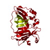
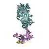
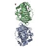
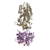
Entry Database : PDB / ID : 4ln9Title Crystal structure of the dehydratase domain from the terminal module of the rifamycin polyketide synthase Rifamycin polyketide synthase Keywords / / / Function / homology Function Domain/homology Component
/ / / / / / / / / / / / / / / / / / / / / / / / / / / / / / / / / / / / / / / / / / / / / / / / / / / / / / / / / / / / / Biological species Amycolatopsis mediterranei (bacteria)Method / / / Resolution : 1.82 Å Authors Gay, D.C. / You, Y.-O. / Cane, D. / Keatinge-Clay, A.T. Journal : Biochemistry / Year : 2013Title : Structure and stereospecificity of the dehydratase domain from the terminal module of the rifamycin polyketide synthase.Authors : Gay, D. / You, Y.O. / Keatinge-Clay, A. / Cane, D.E. History Deposition Jul 11, 2013 Deposition site / Processing site Revision 1.0 Dec 18, 2013 Provider / Type Revision 1.1 Feb 19, 2014 Group Revision 1.2 Sep 20, 2023 Group / Database references / Refinement descriptionCategory chem_comp_atom / chem_comp_bond ... chem_comp_atom / chem_comp_bond / database_2 / pdbx_initial_refinement_model / struct_ref_seq_dif Item / _database_2.pdbx_database_accession / _struct_ref_seq_dif.details
Show all Show less
 Movie
Movie Controller

 Yorodumi
 Open data
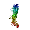
 Basic information
 Components
 Keywords
 Function and homology information
 Amycolatopsis mediterranei (bacteria)
 X-RAY DIFFRACTION /
  Authors
 Citation
 Structure visualization
 Downloads & links
 Other downloads


 PDBj
PDBj


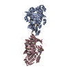
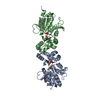
 Assembly
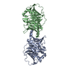
 Mass: 18.015 Da / Num. of mol.: 310 / Source method: isolated from a natural source / Formula: H2O
 Sample preparation
 / Beamline: 5.0.2 / Wavelength: 1.2131 Å
 Processing