 Movie
Movie Controller

[English] 日本語
 Yorodumi
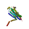
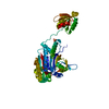
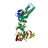
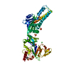
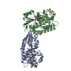
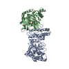
- PDB-5g53: Structure of the adenosine A2A receptor bound to an engineered G ... -
+ Open data

- Basic information
| Entry | Database: PDB / ID: 5g53 | ||||||
|---|---|---|---|---|---|---|---|
| Title | Structure of the adenosine A2A receptor bound to an engineered G protein | ||||||
 Components Components |
| ||||||
 Keywords Keywords | SIGNALING PROTEIN / G PROTEIN COUPLED RECEPTOR / ADENOSINE RECEPTOR / SEVEN-HELIX RECEPTOR / INTEGRAL MEMBRANE PROTEIN / GPCR / ENGINEERED G PROTEIN / GPCR-G PROTEIN COMPLEX / MINI-GS | ||||||
| Function / homology |  Function and homology information Function and homology informationregulation of norepinephrine secretion / negative regulation of alpha-beta T cell activation / positive regulation of circadian sleep/wake cycle, sleep / Adenosine P1 receptors / positive regulation of acetylcholine secretion, neurotransmission / G protein-coupled adenosine receptor activity / sensory perception of chemical stimulus / response to purine-containing compound / mu-type opioid receptor binding / corticotropin-releasing hormone receptor 1 binding ...regulation of norepinephrine secretion / negative regulation of alpha-beta T cell activation / positive regulation of circadian sleep/wake cycle, sleep / Adenosine P1 receptors / positive regulation of acetylcholine secretion, neurotransmission / G protein-coupled adenosine receptor activity / sensory perception of chemical stimulus / response to purine-containing compound / mu-type opioid receptor binding / corticotropin-releasing hormone receptor 1 binding / G protein-coupled adenosine receptor signaling pathway / NGF-independant TRKA activation / Surfactant metabolism / synaptic transmission, dopaminergic / type 5 metabotropic glutamate receptor binding / negative regulation of vascular permeability / beta-2 adrenergic receptor binding / synaptic transmission, cholinergic / intermediate filament / presynaptic active zone / positive regulation of urine volume / response to caffeine / blood circulation / sensory perception / positive regulation of glutamate secretion / eating behavior / inhibitory postsynaptic potential / regulation of calcium ion transport / adenylate cyclase-activating G protein-coupled bile acid receptor signaling pathway / adenylate cyclase-activating serotonin receptor signaling pathway / regulation of skeletal muscle contraction / alpha-actinin binding / PKA activation in glucagon signalling / hair follicle placode formation / developmental growth / intracellular transport / asymmetric synapse / axolemma / membrane depolarization / D1 dopamine receptor binding / cellular defense response / prepulse inhibition / vascular endothelial cell response to laminar fluid shear stress / phagocytosis / renal water homeostasis / Hedgehog 'off' state / activation of adenylate cyclase activity / adenylate cyclase-activating adrenergic receptor signaling pathway / cellular response to acidic pH / insulin-like growth factor receptor binding / positive regulation of synaptic transmission, glutamatergic / neuron projection morphogenesis / cellular response to glucagon stimulus / astrocyte activation / ionotropic glutamate receptor binding / intracellular glucose homeostasis / presynaptic modulation of chemical synaptic transmission / adenylate cyclase activator activity / positive regulation of insulin secretion involved in cellular response to glucose stimulus / trans-Golgi network membrane / positive regulation of long-term synaptic potentiation / positive regulation of synaptic transmission, GABAergic / central nervous system development / positive regulation of protein secretion / response to amphetamine / regulation of mitochondrial membrane potential / positive regulation of apoptotic signaling pathway / negative regulation of inflammatory response to antigenic stimulus / apoptotic signaling pathway / synaptic transmission, glutamatergic / response to prostaglandin E / locomotory behavior / excitatory postsynaptic potential / bone development / platelet aggregation / negative regulation of inflammatory response / cognition / vasodilation / G-protein beta/gamma-subunit complex binding / adenylate cyclase-modulating G protein-coupled receptor signaling pathway / blood coagulation / positive regulation of insulin secretion / sensory perception of smell / Glucagon signaling in metabolic regulation / Prostacyclin signalling through prostacyclin receptor / Glucagon-type ligand receptors / Vasopressin regulates renal water homeostasis via Aquaporins / Glucagon-like Peptide-1 (GLP1) regulates insulin secretion / G alpha (z) signalling events / cellular response to catecholamine stimulus / cell-cell signaling / ADORA2B mediated anti-inflammatory cytokines production / adenylate cyclase-activating dopamine receptor signaling pathway / positive regulation of cold-induced thermogenesis / GPER1 signaling / cellular response to prostaglandin E stimulus / heterotrimeric G-protein complex / adenylate cyclase-activating G protein-coupled receptor signaling pathway / G protein activity / presynaptic membrane Similarity search - Function | ||||||
| Biological species |  HOMO SAPIENS (human) HOMO SAPIENS (human) | ||||||
| Method |  X-RAY DIFFRACTION / SYNCHROTRON / MOLECULAR REPLACEMENT / Resolution: 3.4 Å X-RAY DIFFRACTION / SYNCHROTRON / MOLECULAR REPLACEMENT / Resolution: 3.4 Å | ||||||
 Authors Authors | Carpenter, B. / Nehme, R. / Warne, T. / Leslie, A.G.W. / Tate, C.G. | ||||||
 Citation Citation | Journal: Nature / Year: 2016 Title: Structure of the Adenosine A2A Receptor Bound to an Engineered G Protein Authors: Carpenter, B. / Nehme, R. / Warne, T. / Leslie, A.G.W. / Tate, C.G. #1: Journal: Nature / Year: 2011Title: Agonist-Bound Adenosine A2A Receptor Structures Reveal Common Features of Gpcr Activation Authors: Lebon, G. / Warne, T. / Edwards, P.C. / Bennett, K. / Langmead, C.J. / Leslie, A.G.W. / Tate, C.G. #2: Journal: Protein Eng. Des. Sel. / Year: 2016 Title: Engineering a minimal G protein to facilitate crystallisation of G protein-coupled receptors in their active conformation. Authors: Carpenter, B. / Tate, C.G. | ||||||
| History |
|
- Structure visualization
| Structure viewer | Molecule: MolmilJmol/JSmol |
|---|
- Downloads & links
-Download
| PDBx/mmCIF format | 5g53.cif.gz | 198.9 KB | Display | PDBx/mmCIF format |
|---|---|---|---|---|
| PDB format | pdb5g53.ent.gz | 155.5 KB | Display | PDB format |
| PDBx/mmJSON format | 5g53.json.gz | Tree view | PDBx/mmJSON format | |
| Others |  Other downloads Other downloads |
-Validation report
| Arichive directory | https://data.pdbj.org/pub/pdb/validation_reports/g5/5g53ftp://data.pdbj.org/pub/pdb/validation_reports/g5/5g53 | HTTPS FTP |
|---|
-Related structure data


-Links
 PDBj
PDBj


- Assembly
| Deposited unit | 
| ||||||||
|---|---|---|---|---|---|---|---|---|---|
| 1 | 
| ||||||||
| 2 | 
| ||||||||
| Unit cell |
|
-Components
| #1: Protein | Mass: 34909.500 Da / Num. of mol.: 2 / Mutation: YES Source method: isolated from a genetically manipulated source Details: THE CONSTRUCT WAS TRUNCATED AFTER RESIDUE 308 OF THE A2A SEQUENCE, BUT HAS A 6 RESIDUE SEQUENCE THAT INCLUDES THE TEV CLEAVAGE SEQUENCE (ENLYFQ) AT THE C- TERMINUS Source: (gene. exp.) HOMO SAPIENS (human) / Tissue: BRAIN / Plasmid: PBACPAK8 / Production host:  TRICHOPLUSIA NI (cabbage looper) / References: UniProt: P29274 TRICHOPLUSIA NI (cabbage looper) / References: UniProt: P29274#2: Protein | Mass: 26625.125 Da / Num. of mol.: 2 / Fragment: RAS DOMAIN, RESIDUES 26-60 AND 847-1037 / Mutation: YES Source method: isolated from a genetically manipulated source Details: DELETIONS 1-25,65-203,255-264 INSERTION GGSGGSGG LINKING RESIDUES 64 AND 204 Source: (gene. exp.) HOMO SAPIENS (human) / Tissue: BRAIN / Production host:  #3: Chemical | 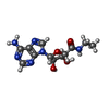  Mass: 308.293 Da / Num. of mol.: 2 / Source method: obtained synthetically / Formula: C12H16N6O4 Mass: 308.293 Da / Num. of mol.: 2 / Source method: obtained synthetically / Formula: C12H16N6O4#4: Sugar | 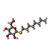  Type: D-saccharide / Mass: 308.434 Da / Num. of mol.: 2 Type: D-saccharide / Mass: 308.434 Da / Num. of mol.: 2Source method: isolated from a genetically manipulated source Formula: C14H28O5S / Comment: detergent*YM #5: Chemical | ChemComp-GDP / |   Type: RNA linking / Mass: 443.201 Da / Num. of mol.: 1 / Source method: obtained synthetically / Formula: C10H15N5O11P2 / Comment: GDP, energy-carrying molecule*YM Type: RNA linking / Mass: 443.201 Da / Num. of mol.: 1 / Source method: obtained synthetically / Formula: C10H15N5O11P2 / Comment: GDP, energy-carrying molecule*YMHas protein modification | Y | Sequence details | A2A RECEPTOR. THE CONSTRUCT WAS TRUNCATED AFTER RESIDUE 308 OF THE A2A SEQUENCE. REMOVAL OF ...A2A RECEPTOR. THE CONSTRUCT WAS TRUNCATED AFTER RESIDUE 308 OF THE A2A SEQUENCE. REMOVAL OF GLYCOSYLAT | |
|---|
-Experimental details
-Experiment
| Experiment | Method: X-RAY DIFFRACTION / Number of used crystals: 2 |
|---|
- Sample preparation
| Crystal | Density Matthews: 2.9 Å3/Da / Density % sol: 58 % Description: THE RAS DOMAIN FROM ENTRY 3SN6 WAS USED FOR MOLECULAR REPLACEMENT |
|---|---|
| Crystal grow | pH: 5.5 Details: 0.1 M NAOAC PH 5.5, 10% PEG 2000 (IN THE PRESENCE OF CHS); OR 0.1 M NAOAC PH 5.7, 9.5% PEG 2000 MME (IN THE ABSENCE OF CHS) |
-Data collection
| Diffraction | Mean temperature: 100 K |
|---|---|
| Diffraction source | Source: SYNCHROTRON / Site: ESRF  / Beamline: ID23-2 / Wavelength: 0.8729 / Beamline: ID23-2 / Wavelength: 0.8729 |
| Detector | Type: DECTRIS PILATUS 2M / Detector: PIXEL / Date: Apr 9, 2015 / Details: KB MIRRORS |
| Radiation | Monochromator: SI(111) / Protocol: SINGLE WAVELENGTH / Monochromatic (M) / Laue (L): M / Scattering type: x-ray |
| Radiation wavelength | Wavelength: 0.8729 Å / Relative weight: 1 |
| Reflection | Resolution: 3.4→40.3 Å / Num. obs: 20898 / % possible obs: 90.6 % / Observed criterion σ(I): 0 / Redundancy: 2.6 % / Rmerge(I) obs: 0.17 / Net I/σ(I): 3.6 |
| Reflection shell | Resolution: 3.4→3.49 Å / Redundancy: 2.4 % / Rmerge(I) obs: 0.75 / Mean I/σ(I) obs: 1.2 / % possible all: 78.5 |
- Processing
| Software |
| ||||||||||||||||||||||||||||||||||||||||||||||||||||||||||||||||||||||||||||||||||||||||||||||||||||||||||||||||||||||||||||||||||||||||||||||||||||||||||||||||||||||||||||||||||||||
|---|---|---|---|---|---|---|---|---|---|---|---|---|---|---|---|---|---|---|---|---|---|---|---|---|---|---|---|---|---|---|---|---|---|---|---|---|---|---|---|---|---|---|---|---|---|---|---|---|---|---|---|---|---|---|---|---|---|---|---|---|---|---|---|---|---|---|---|---|---|---|---|---|---|---|---|---|---|---|---|---|---|---|---|---|---|---|---|---|---|---|---|---|---|---|---|---|---|---|---|---|---|---|---|---|---|---|---|---|---|---|---|---|---|---|---|---|---|---|---|---|---|---|---|---|---|---|---|---|---|---|---|---|---|---|---|---|---|---|---|---|---|---|---|---|---|---|---|---|---|---|---|---|---|---|---|---|---|---|---|---|---|---|---|---|---|---|---|---|---|---|---|---|---|---|---|---|---|---|---|---|---|---|---|
| Refinement | Method to determine structure: MOLECULAR REPLACEMENT Starting model: PDB ENTRIES 2YDV, 3SN6 Resolution: 3.4→91.89 Å / Cor.coef. Fo:Fc: 0.828 / Cor.coef. Fo:Fc free: 0.811 / SU B: 46.428 / SU ML: 0.697 / Cross valid method: THROUGHOUT / ESU R Free: 0.69 / Stereochemistry target values: MAXIMUM LIKELIHOOD Details: HYDROGENS HAVE BEEN ADDED IN THE RIDING POSITIONS. U VALUES REFINED INDIVIDUALLY SIDE CHAINS WITHOUT CLEAR ELECTRON DENSITY HAVE BEEN TRUNCATED BACK TO CBETA.
| ||||||||||||||||||||||||||||||||||||||||||||||||||||||||||||||||||||||||||||||||||||||||||||||||||||||||||||||||||||||||||||||||||||||||||||||||||||||||||||||||||||||||||||||||||||||
| Solvent computation | Ion probe radii: 0.8 Å / Shrinkage radii: 0.8 Å / VDW probe radii: 1.2 Å / Solvent model: MASK | ||||||||||||||||||||||||||||||||||||||||||||||||||||||||||||||||||||||||||||||||||||||||||||||||||||||||||||||||||||||||||||||||||||||||||||||||||||||||||||||||||||||||||||||||||||||
| Displacement parameters | Biso mean: 76.297 Å2
| ||||||||||||||||||||||||||||||||||||||||||||||||||||||||||||||||||||||||||||||||||||||||||||||||||||||||||||||||||||||||||||||||||||||||||||||||||||||||||||||||||||||||||||||||||||||
| Refinement step | Cycle: LAST / Resolution: 3.4→91.89 Å
| ||||||||||||||||||||||||||||||||||||||||||||||||||||||||||||||||||||||||||||||||||||||||||||||||||||||||||||||||||||||||||||||||||||||||||||||||||||||||||||||||||||||||||||||||||||||
| Refine LS restraints |
|