 Movie
Movie Controller


+ Open data

- Basic information
| Entry | Database: PDB / ID: 6m3i | ||||||
|---|---|---|---|---|---|---|---|
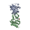
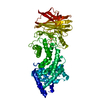
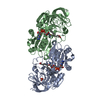
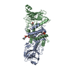
| Title | Crystal structure of HPF1/PARP1 complex | ||||||
 Components Components |
| ||||||
 Keywords Keywords | NUCLEAR PROTEIN / complex / ADP-ribosylation / DNA damage response | ||||||
| Function / homology |  Function and homology information Function and homology informationregulation of protein ADP-ribosylation / protein ADP-ribosyltransferase-substrate adaptor activity / NAD+-histone H2BS6 serine ADP-ribosyltransferase activity / NAD+-histone H3S10 serine ADP-ribosyltransferase activity / NAD+-histone H2BE35 glutamate ADP-ribosyltransferase activity / positive regulation of myofibroblast differentiation / negative regulation of ATP biosynthetic process / poly-ADP-D-ribose binding / NAD+-protein-tyrosine ADP-ribosyltransferase activity / NAD+-protein-histidine ADP-ribosyltransferase activity ...regulation of protein ADP-ribosylation / protein ADP-ribosyltransferase-substrate adaptor activity / NAD+-histone H2BS6 serine ADP-ribosyltransferase activity / NAD+-histone H3S10 serine ADP-ribosyltransferase activity / NAD+-histone H2BE35 glutamate ADP-ribosyltransferase activity / positive regulation of myofibroblast differentiation / negative regulation of ATP biosynthetic process / poly-ADP-D-ribose binding / NAD+-protein-tyrosine ADP-ribosyltransferase activity / NAD+-protein-histidine ADP-ribosyltransferase activity / non-sequence-specific DNA binding, bending / regulation of base-excision repair / regulation of circadian sleep/wake cycle, non-REM sleep / mitochondrial DNA metabolic process / vRNA Synthesis / single-strand break-containing DNA binding / carbohydrate biosynthetic process / NAD+-protein-serine ADP-ribosyltransferase activity / NAD DNA ADP-ribosyltransferase activity / negative regulation of adipose tissue development / DNA ADP-ribosylation / regulation of oxidative stress-induced neuron intrinsic apoptotic signaling pathway / ATP generation from poly-ADP-D-ribose / replication fork reversal / establishment of protein localization to chromatin / positive regulation of necroptotic process / signal transduction involved in regulation of gene expression / transcription regulator activator activity / response to aldosterone / HDR through MMEJ (alt-NHEJ) / single strand break repair / positive regulation of intracellular estrogen receptor signaling pathway / positive regulation of DNA-templated transcription, elongation / NAD+ ADP-ribosyltransferase / protein auto-ADP-ribosylation / negative regulation of telomere maintenance via telomere lengthening / NAD+-protein-aspartate ADP-ribosyltransferase activity / mitochondrial DNA repair / protein poly-ADP-ribosylation / NAD+-protein-glutamate ADP-ribosyltransferase activity / positive regulation of cardiac muscle hypertrophy / negative regulation of cGAS/STING signaling pathway / NAD+-protein mono-ADP-ribosyltransferase activity / decidualization / cellular response to zinc ion / protein autoprocessing / positive regulation of mitochondrial depolarization / negative regulation of transcription elongation by RNA polymerase II / R-SMAD binding / macrophage differentiation / nuclear replication fork / Transferases; Glycosyltransferases; Pentosyltransferases / positive regulation of SMAD protein signal transduction / POLB-Dependent Long Patch Base Excision Repair / NAD+ poly-ADP-ribosyltransferase activity / DNA repair-dependent chromatin remodeling / SUMOylation of DNA damage response and repair proteins / nucleosome binding / positive regulation of double-strand break repair via homologous recombination / site of DNA damage / protein localization to chromatin / nucleotidyltransferase activity / transforming growth factor beta receptor signaling pathway / negative regulation of innate immune response / telomere maintenance / nuclear estrogen receptor binding / protein modification process / response to gamma radiation / mitochondrion organization / Downregulation of SMAD2/3:SMAD4 transcriptional activity / cellular response to nerve growth factor stimulus / protein-DNA complex / positive regulation of protein localization to nucleus / fibrillar center / DNA Damage Recognition in GG-NER / NAD binding / cellular response to amyloid-beta / histone deacetylase binding / enzyme activator activity / Dual Incision in GG-NER / cellular response to insulin stimulus / Formation of Incision Complex in GG-NER / cellular response to UV / nuclear envelope / double-strand break repair / regulation of protein localization / site of double-strand break / cellular response to oxidative stress / transcription by RNA polymerase II / transcription regulator complex / histone binding / damaged DNA binding / RNA polymerase II-specific DNA-binding transcription factor binding / response to ethanol / positive regulation of canonical NF-kappaB signal transduction / chromosome, telomeric region / nuclear body / innate immune response / negative regulation of DNA-templated transcription / DNA repair Similarity search - Function | ||||||
| Biological species |  Homo sapiens (human) Homo sapiens (human) | ||||||
| Method |  X-RAY DIFFRACTION / SYNCHROTRON / MOLECULAR REPLACEMENT / Resolution: 1.98 Å X-RAY DIFFRACTION / SYNCHROTRON / MOLECULAR REPLACEMENT / Resolution: 1.98 Å | ||||||
 Authors Authors | Sun, F.H. / Yun, C.H. | ||||||
| Funding support |  China, 1items China, 1items
| ||||||
 Citation Citation | Journal: Nat Commun / Year: 2021 Title: HPF1 remodels the active site of PARP1 to enable the serine ADP-ribosylation of histones. Authors: Sun, F.H. / Zhao, P. / Zhang, N. / Kong, L.L. / Wong, C.C.L. / Yun, C.H. | ||||||
| History |
|
- Structure visualization
| Structure viewer | Molecule: MolmilJmol/JSmol |
|---|
- Downloads & links
-Download
| PDBx/mmCIF format | 6m3i.cif.gz | 143.5 KB | Display | PDBx/mmCIF format |
|---|---|---|---|---|
| PDB format | pdb6m3i.ent.gz | 99.2 KB | Display | PDB format |
| PDBx/mmJSON format | 6m3i.json.gz | Tree view | PDBx/mmJSON format | |
| Others |  Other downloads Other downloads |
-Validation report
| Arichive directory | https://data.pdbj.org/pub/pdb/validation_reports/m3/6m3iftp://data.pdbj.org/pub/pdb/validation_reports/m3/6m3i | HTTPS FTP |
|---|
-Related structure data
| Related structure data | 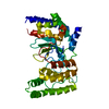 6m3gSC 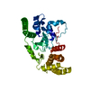 6m3hC  6bhvS S: Starting model for refinement C: citing same article ( |
|---|---|
| Similar structure data |


-Links
 PDBj
PDBj


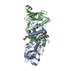
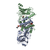
- Assembly
| Deposited unit | 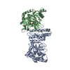
| ||||||||||||
|---|---|---|---|---|---|---|---|---|---|---|---|---|---|
| 1 |
| ||||||||||||
| Unit cell |
|
-Components
| #1: Protein | Mass: 39495.082 Da / Num. of mol.: 1 Source method: isolated from a genetically manipulated source Source: (gene. exp.) Homo sapiens (human) / Gene: HPF1, C4orf27Production host: References: UniProt: Q9NWY4 |
|---|---|
| #2: Protein | Mass: 28057.139 Da / Num. of mol.: 1 Source method: isolated from a genetically manipulated source Source: (gene. exp.) Homo sapiens (human) / Gene: PARP1, ADPRT, PPOLProduction host: References: UniProt: P09874, NAD+ ADP-ribosyltransferase, Transferases; Glycosyltransferases; Pentosyltransferases |
| #3: Chemical | ChemComp-UNU /   Mass: 121.137 Da / Num. of mol.: 1 / Source method: obtained synthetically / Formula: C7H7NO / Feature type: SUBJECT OF INVESTIGATION Mass: 121.137 Da / Num. of mol.: 1 / Source method: obtained synthetically / Formula: C7H7NO / Feature type: SUBJECT OF INVESTIGATION |
| #4: Water | ChemComp-HOH /  Mass: 18.015 Da / Num. of mol.: 323 / Source method: isolated from a natural source / Formula: H2O Mass: 18.015 Da / Num. of mol.: 323 / Source method: isolated from a natural source / Formula: H2O |
| Has ligand of interest | Y |
-Experimental details
-Experiment
| Experiment | Method: X-RAY DIFFRACTION / Number of used crystals: 1 |
|---|
- Sample preparation
| Crystal | Density Matthews: 2.42 Å3/Da / Density % sol: 49.21 % |
|---|---|
| Crystal grow | Temperature: 300 K / Method: vapor diffusion Details: 0.1 M Tris-pH 7.0, 0.2 M magnesium formate dehydrate, 20% w/v PEG 3350. |
-Data collection
| Diffraction | Mean temperature: 100 K / Serial crystal experiment: N |
|---|---|
| Diffraction source | Source: SYNCHROTRON / Site: SSRF / Beamline: BL19U1 / Wavelength: 0.97849 Å |
| Detector | Type: Nonius Kappa CCD / Detector: CCD / Date: Feb 16, 2019 |
| Radiation | Protocol: SINGLE WAVELENGTH / Monochromatic (M) / Laue (L): M / Scattering type: x-ray |
| Radiation wavelength | Wavelength: 0.97849 Å / Relative weight: 1 |
| Reflection | Resolution: 1.98→50 Å / Num. obs: 44217 / % possible obs: 98.3 % / Redundancy: 3.4 % / Biso Wilson estimate: 27.82 Å2 / Rpim(I) all: 0.053 / Net I/σ(I): 14.1 |
| Reflection shell | Resolution: 1.98→2.03 Å / Num. unique obs: 2414 / Rpim(I) all: 0.385 |
- Processing
| Software |
| |||||||||||||||||||||||||||||||||||||||||||||||||||||||||||||||||||||||||||||||||||||||||||||||||||||||||||||||||||||||
|---|---|---|---|---|---|---|---|---|---|---|---|---|---|---|---|---|---|---|---|---|---|---|---|---|---|---|---|---|---|---|---|---|---|---|---|---|---|---|---|---|---|---|---|---|---|---|---|---|---|---|---|---|---|---|---|---|---|---|---|---|---|---|---|---|---|---|---|---|---|---|---|---|---|---|---|---|---|---|---|---|---|---|---|---|---|---|---|---|---|---|---|---|---|---|---|---|---|---|---|---|---|---|---|---|---|---|---|---|---|---|---|---|---|---|---|---|---|---|---|---|
| Refinement | Method to determine structure: MOLECULAR REPLACEMENT Starting model: 6M3G, 6BHV Resolution: 1.98→27.39 Å / SU ML: 0.2247 / Cross valid method: NONE / σ(F): 1.36 / Phase error: 21.1342
| |||||||||||||||||||||||||||||||||||||||||||||||||||||||||||||||||||||||||||||||||||||||||||||||||||||||||||||||||||||||
| Solvent computation | Shrinkage radii: 0.9 Å / VDW probe radii: 1.11 Å | |||||||||||||||||||||||||||||||||||||||||||||||||||||||||||||||||||||||||||||||||||||||||||||||||||||||||||||||||||||||
| Displacement parameters | Biso mean: 31.03 Å2 | |||||||||||||||||||||||||||||||||||||||||||||||||||||||||||||||||||||||||||||||||||||||||||||||||||||||||||||||||||||||
| Refinement step | Cycle: LAST / Resolution: 1.98→27.39 Å
| |||||||||||||||||||||||||||||||||||||||||||||||||||||||||||||||||||||||||||||||||||||||||||||||||||||||||||||||||||||||
| Refine LS restraints |
| |||||||||||||||||||||||||||||||||||||||||||||||||||||||||||||||||||||||||||||||||||||||||||||||||||||||||||||||||||||||
| LS refinement shell |
|
