 Movie
Movie Controller

[English] 日本語
 Yorodumi
- PDB-1dlq: STRUCTURE OF CATECHOL 1,2-DIOXYGENASE FROM ACINETOBACTER SP. ADP1... -
+ Open data

- Basic information
| Entry | Database: PDB / ID: 1dlq | ||||||
|---|---|---|---|---|---|---|---|
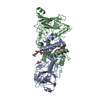
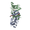
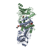
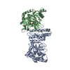
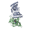
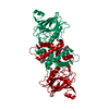
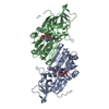
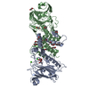
| Title | STRUCTURE OF CATECHOL 1,2-DIOXYGENASE FROM ACINETOBACTER SP. ADP1 INHIBITED BY BOUND MERCURY | ||||||
 Components Components | CATECHOL 1,2-DIOXYGENASE | ||||||
 Keywords Keywords | OXIDOREDUCTASE / METALLOPROTEIN / DIOXYGENASE / MERCURY / AROMATIC COMPOUND DEGREDATION / MIXED ALPHA/BETA STRUCTURE | ||||||
| Function / homology |  Function and homology information Function and homology informationcatechol-containing compound catabolic process / catechol 1,2-dioxygenase / catechol 1,2-dioxygenase activity / beta-ketoadipate pathway / ferric iron binding Similarity search - Function | ||||||
| Biological species |  Acinetobacter sp. (bacteria) Acinetobacter sp. (bacteria) | ||||||
| Method |  X-RAY DIFFRACTION / Resolution: 2.3 Å X-RAY DIFFRACTION / Resolution: 2.3 Å | ||||||
 Authors Authors | Vetting, M.W. / Ohlendorf, D.H. | ||||||
 Citation Citation | Journal: Structure Fold.Des. / Year: 2000 Title: The 1.8 A crystal structure of catechol 1,2-dioxygenase reveals a novel hydrophobic helical zipper as a subunit linker. Authors: Vetting, M.W. / Ohlendorf, D.H. #1: Journal: J.Bacteriol. / Year: 1988Title: DNA Sequence of the Acinetobacter calcoaceticus Catechol 1,2-dioxygenase I Structural Gene catA: Evidence for Evolutionary Divergence of Intradiol Dioxygenases by Aquisition of DNA Sequence Repetitions Authors: Neidle, E.L. / Harnett, C. / Bonitz, S. / Ornston, L.N. #2: Journal: J.Mol.Biol. / Year: 1994Title: Structure of Protocatechuate 3,4-dioxygenase from Psuedomonas aeruginosa at 2.15 A Resolution Authors: Ohlendorf, D.H. / Orville, A.M. / Lipscomb, J.D. #3: Journal: Biochemistry / Year: 1997Title: Crystal Structures of Substrate and Substrate Analog Complexes of Protocatechuate 3,4-dioxygenase: Endogenous Fe+3 Ligand Displacement in Response to Substrate Binding. Authors: Orville, A.M. / Lipscomb, J.D. / Ohlendorf, D.H. | ||||||
| History |
|
- Structure visualization
| Structure viewer | Molecule: MolmilJmol/JSmol |
|---|
- Downloads & links
-Download
| PDBx/mmCIF format | 1dlq.cif.gz | 135.6 KB | Display | PDBx/mmCIF format |
|---|---|---|---|---|
| PDB format | pdb1dlq.ent.gz | 106 KB | Display | PDB format |
| PDBx/mmJSON format | 1dlq.json.gz | Tree view | PDBx/mmJSON format | |
| Others |  Other downloads Other downloads |
-Validation report
| Arichive directory | https://data.pdbj.org/pub/pdb/validation_reports/dl/1dlqftp://data.pdbj.org/pub/pdb/validation_reports/dl/1dlq | HTTPS FTP |
|---|
-Related structure data


-Links
 PDBj
PDBj


- Assembly
| Deposited unit | 
| ||||||||
|---|---|---|---|---|---|---|---|---|---|
| 1 |
| ||||||||
| Unit cell |
| ||||||||
| Details | The biological assembly of the molecule is a homodimer consisting of subunit A and subunit B related by a non-crystallographic two-fold |
-Components
| #1: Protein | Mass: 34384.145 Da / Num. of mol.: 2 Source method: isolated from a genetically manipulated source Source: (gene. exp.) Acinetobacter sp. (bacteria) / Strain: ADP1 / Production host: #2: Chemical |   Mass: 55.845 Da / Num. of mol.: 2 / Source method: obtained synthetically / Formula: Fe Mass: 55.845 Da / Num. of mol.: 2 / Source method: obtained synthetically / Formula: Fe#3: Chemical | ChemComp-HG /   Mass: 200.590 Da / Num. of mol.: 6 / Source method: obtained synthetically / Formula: Hg Mass: 200.590 Da / Num. of mol.: 6 / Source method: obtained synthetically / Formula: Hg#4: Chemical |   Mass: 636.861 Da / Num. of mol.: 2 / Source method: obtained synthetically / Formula: C33H67NO8P Mass: 636.861 Da / Num. of mol.: 2 / Source method: obtained synthetically / Formula: C33H67NO8P#5: Water | ChemComp-HOH / |  Mass: 18.015 Da / Num. of mol.: 125 / Source method: isolated from a natural source / Formula: H2O Mass: 18.015 Da / Num. of mol.: 125 / Source method: isolated from a natural source / Formula: H2O |
|---|
-Experimental details
-Experiment
| Experiment | Method: X-RAY DIFFRACTION / Number of used crystals: 1 |
|---|
- Sample preparation
| Crystal | Density Matthews: 2.8 Å3/Da / Density % sol: 56.07 % | |||||||||||||||||||||||||
|---|---|---|---|---|---|---|---|---|---|---|---|---|---|---|---|---|---|---|---|---|---|---|---|---|---|---|
| Crystal grow | Temperature: 277 K / Method: vapor diffusion, hanging drop / pH: 7.5 Details: 10-15% Peg5000, 100mM Tris-HCl, pH 7.5, .2M MgAcetate , VAPOR DIFFUSION, HANGING DROP, temperature 277K | |||||||||||||||||||||||||
| Crystal grow | *PLUS Temperature: 4 ℃ | |||||||||||||||||||||||||
| Components of the solutions | *PLUS
|
-Data collection
| Diffraction | Mean temperature: 298 K |
|---|---|
| Diffraction source | Source: ROTATING ANODE / Type: RIGAKU RU200 / Wavelength: 1.54 |
| Detector | Type: SIEMENS HI-STAR / Detector: AREA DETECTOR / Date: Sep 22, 1998 |
| Radiation | Protocol: SINGLE WAVELENGTH / Monochromatic (M) / Laue (L): M / Scattering type: x-ray |
| Radiation wavelength | Wavelength: 1.54 Å / Relative weight: 1 |
| Reflection | Resolution: 2.3→100 Å / Num. all: 30521 / Num. obs: 76231 / % possible obs: 90.7 % / Observed criterion σ(F): 0 / Observed criterion σ(I): 0 / Redundancy: 2.5 % / Biso Wilson estimate: 33.9 Å2 / Rmerge(I) obs: 0.037 / Net I/σ(I): 31 |
| Reflection shell | Resolution: 2.31→2.41 Å / Redundancy: 1.48 % / Rmerge(I) obs: 0.118 / Num. unique all: 2550 / % possible all: 46 |
| Reflection | *PLUS Num. obs: 33625 / Num. measured all: 76231 / Rmerge(I) obs: 0.038 |
| Reflection shell | *PLUS % possible obs: 45.9 % / Mean I/σ(I) obs: 5 |
- Processing
| Software |
| |||||||||||||||||||||||||
|---|---|---|---|---|---|---|---|---|---|---|---|---|---|---|---|---|---|---|---|---|---|---|---|---|---|---|
| Refinement | Resolution: 2.3→20 Å / σ(F): 0 / σ(I): 0 / Stereochemistry target values: Engh & Huber
| |||||||||||||||||||||||||
| Refinement step | Cycle: LAST / Resolution: 2.3→20 Å
| |||||||||||||||||||||||||
| Refine LS restraints |
| |||||||||||||||||||||||||
| Software | *PLUS Name: CNS / Classification: refinement | |||||||||||||||||||||||||
| Refinement | *PLUS Highest resolution: 2.3 Å / Lowest resolution: 20 Å / σ(F): 0 / % reflection Rfree: 10 % / Rfactor obs: 0.182 | |||||||||||||||||||||||||
| Solvent computation | *PLUS | |||||||||||||||||||||||||
| Displacement parameters | *PLUS | |||||||||||||||||||||||||
| Refine LS restraints | *PLUS Type: c_angle_deg / Dev ideal: 1.5 |
