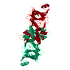
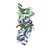
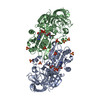
Entry Database : PDB / ID : 2xsvTitle Crystal structure of L69A mutant Acinetobacter radioresistens catechol 1,2 dioxygenase CATECHOL 1,2 DIOXYGENASE Keywords Function / homology Function Domain/homology Component
/ / / / / / / / / / / / / / / / / / / / / / / / Biological species ACINETOBACTER RADIORESISTENS (bacteria)Method / / / Resolution : 1.8 Å Authors Micalella, C. / Martignon, S. / Bruno, S. / Rizzi, M. Journal : Biochim.Biophys.Acta / Year : 2011Title : X-Ray Crystallography, Mass Spectrometry and Single Crystal Microspectrophotometry: A Multidisciplinary Characterization of Catechol 1,2 Dioxygenase.Authors : Micalella, C. / Martignon, S. / Bruno, S. / Pioselli, B. / Caglio, R. / Valetti, F. / Pessione, E. / Giunta, C. / Rizzi, M. History Deposition Sep 30, 2010 Deposition site / Processing site Revision 1.0 Oct 13, 2010 Provider / Type Revision 1.1 Dec 18, 2013 Group / Structure summary / Version format complianceRevision 1.2 Dec 20, 2023 Group Data collection / Database references ... Data collection / Database references / Derived calculations / Other / Refinement description Category chem_comp_atom / chem_comp_bond ... chem_comp_atom / chem_comp_bond / database_2 / pdbx_database_status / pdbx_initial_refinement_model / pdbx_struct_conn_angle / struct_conn / struct_site Item _database_2.pdbx_DOI / _database_2.pdbx_database_accession ... _database_2.pdbx_DOI / _database_2.pdbx_database_accession / _pdbx_database_status.status_code_sf / _pdbx_struct_conn_angle.ptnr1_auth_comp_id / _pdbx_struct_conn_angle.ptnr1_auth_seq_id / _pdbx_struct_conn_angle.ptnr1_label_asym_id / _pdbx_struct_conn_angle.ptnr1_label_atom_id / _pdbx_struct_conn_angle.ptnr1_label_comp_id / _pdbx_struct_conn_angle.ptnr1_label_seq_id / _pdbx_struct_conn_angle.ptnr3_auth_comp_id / _pdbx_struct_conn_angle.ptnr3_auth_seq_id / _pdbx_struct_conn_angle.ptnr3_label_asym_id / _pdbx_struct_conn_angle.ptnr3_label_atom_id / _pdbx_struct_conn_angle.ptnr3_label_comp_id / _pdbx_struct_conn_angle.ptnr3_label_seq_id / _pdbx_struct_conn_angle.value / _struct_conn.pdbx_dist_value / _struct_conn.ptnr1_auth_comp_id / _struct_conn.ptnr1_auth_seq_id / _struct_conn.ptnr1_label_asym_id / _struct_conn.ptnr1_label_atom_id / _struct_conn.ptnr1_label_comp_id / _struct_conn.ptnr1_label_seq_id / _struct_conn.ptnr2_auth_comp_id / _struct_conn.ptnr2_auth_seq_id / _struct_conn.ptnr2_label_asym_id / _struct_conn.ptnr2_label_atom_id / _struct_conn.ptnr2_label_comp_id / _struct_conn.ptnr2_label_seq_id / _struct_site.pdbx_auth_asym_id / _struct_site.pdbx_auth_comp_id / _struct_site.pdbx_auth_seq_id Revision 1.3 Dec 24, 2025 Group / Structure summaryCategory chem_comp / entity ... chem_comp / entity / pdbx_entity_nonpoly / pdbx_entry_details Item _chem_comp.name / _entity.pdbx_description ... _chem_comp.name / _entity.pdbx_description / _pdbx_entity_nonpoly.name / _pdbx_entry_details.has_protein_modification
Show all Show less
 Movie
Movie Controller

 Yorodumi
 Open data
 Basic information
 Components
 Keywords
 Function and homology information
 ACINETOBACTER RADIORESISTENS (bacteria)
 X-RAY DIFFRACTION /
  Authors
 Citation
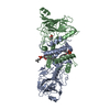
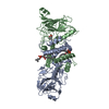
 Structure visualization
 Downloads & links
 Other downloads


 PDBj
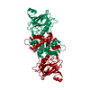
PDBj Assembly
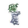
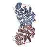


 Mass: 55.845 Da / Num. of mol.: 1 / Source method: obtained synthetically / Formula: Fe
 Mass: 836.061 Da / Num. of mol.: 1 / Source method: obtained synthetically / Formula: C43H80O13P
 Mass: 18.015 Da / Num. of mol.: 290 / Source method: isolated from a natural source / Formula: H2O
 Sample preparation
 / Beamline: ID14-1 / Wavelength: 0.981
  Processing