 Movie
Movie Controller

[English] 日本語
 Yorodumi
- PDB-2xsr: Crystal structure of wild type Acinetobacter radioresistens catec... -
+ Open data

- Basic information
| Entry | Database: PDB / ID: 2xsr | ||||||
|---|---|---|---|---|---|---|---|
| Title | Crystal structure of wild type Acinetobacter radioresistens catechol 1,2 dioxygenase | ||||||
 Components Components | CATECHOL 1,2 DIOXYGENASE | ||||||
 Keywords Keywords | OXIDOREDUCTASE / LIPID | ||||||
| Function / homology |  Function and homology information Function and homology informationcatechol-containing compound catabolic process / catechol 1,2-dioxygenase / catechol 1,2-dioxygenase activity / beta-ketoadipate pathway / ferric iron binding Similarity search - Function | ||||||
| Biological species |  ACINETOBACTER RADIORESISTENS (bacteria) ACINETOBACTER RADIORESISTENS (bacteria) | ||||||
| Method |  X-RAY DIFFRACTION / SYNCHROTRON / MOLECULAR REPLACEMENT / Resolution: 1.8 Å X-RAY DIFFRACTION / SYNCHROTRON / MOLECULAR REPLACEMENT / Resolution: 1.8 Å | ||||||
 Authors Authors | Micalella, C. / Martignon, S. / Bruno, S. / Rizzi, M. | ||||||
 Citation Citation | Journal: Biochim.Biophys.Acta / Year: 2011 Title: X-Ray Crystallography, Mass Spectrometry and Single Crystal Microspectrophotometry: A Multidisciplinary Characterization of Catechol 1,2 Dioxygenase. Authors: Micalella, C. / Martignon, S. / Bruno, S. / Pioselli, B. / Caglio, R. / Valetti, F. / Pessione, E. / Giunta, C. / Rizzi, M. | ||||||
| History |
|
- Structure visualization
| Structure viewer | Molecule: MolmilJmol/JSmol |
|---|
- Downloads & links
-Download
| PDBx/mmCIF format | 2xsr.cif.gz | 83 KB | Display | PDBx/mmCIF format |
|---|---|---|---|---|
| PDB format | pdb2xsr.ent.gz | 60.5 KB | Display | PDB format |
| PDBx/mmJSON format | 2xsr.json.gz | Tree view | PDBx/mmJSON format | |
| Others |  Other downloads Other downloads |
-Validation report
| Arichive directory | https://data.pdbj.org/pub/pdb/validation_reports/xs/2xsrftp://data.pdbj.org/pub/pdb/validation_reports/xs/2xsr | HTTPS FTP |
|---|
-Related structure data
| Related structure data |  2xsuC  2xsvC  2azqS C: citing same article ( S: Starting model for refinement |
|---|---|
| Similar structure data |


-Links
 PDBj
PDBj- Assembly
| Deposited unit | 
| |||||||||
|---|---|---|---|---|---|---|---|---|---|---|
| 1 | 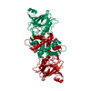
| |||||||||
| Unit cell |
| |||||||||
| Components on special symmetry positions |
|
-Components
| #1: Protein | Mass: 34412.188 Da / Num. of mol.: 1 / Fragment: RESIDUES 2-306 Source method: isolated from a genetically manipulated source Source: (gene. exp.) ACINETOBACTER RADIORESISTENS (bacteria)Strain: LMG S13 / Production host: |
|---|---|
| #2: Chemical | ChemComp-FE /   Mass: 55.845 Da / Num. of mol.: 1 / Source method: obtained synthetically / Formula: Fe Mass: 55.845 Da / Num. of mol.: 1 / Source method: obtained synthetically / Formula: Fe |
| #3: Chemical | ChemComp-PIE /   Mass: 836.061 Da / Num. of mol.: 1 / Source method: obtained synthetically / Formula: C43H80O13P Mass: 836.061 Da / Num. of mol.: 1 / Source method: obtained synthetically / Formula: C43H80O13P |
| #4: Water | ChemComp-HOH /  Mass: 18.015 Da / Num. of mol.: 270 / Source method: isolated from a natural source / Formula: H2O Mass: 18.015 Da / Num. of mol.: 270 / Source method: isolated from a natural source / Formula: H2O |
| Has protein modification | N |
| Nonpolymer details | PHOSHOLIPI |
-Experimental details
-Experiment
| Experiment | Method: X-RAY DIFFRACTION |
|---|
- Sample preparation
| Crystal | Density Matthews: 3.61 Å3/Da / Density % sol: 65.96 % / Description: NONE |
|---|---|
| Crystal grow | Temperature: 277 K Details: 2 M AMMONIUM SULPHATE, 0.1 M TRIS- HCL PH 8.5, 277K. |
-Data collection
| Diffraction | Mean temperature: 100 K |
|---|---|
| Diffraction source | Source: SYNCHROTRON / Site: ESRF  / Beamline: ID14-1 / Wavelength: 0.981 / Beamline: ID14-1 / Wavelength: 0.981 |
| Detector | Type: ADSC QUANTUM 210 / Detector: CCD |
| Radiation | Protocol: SINGLE WAVELENGTH / Monochromatic (M) / Laue (L): M / Scattering type: x-ray |
| Radiation wavelength | Wavelength: 0.981 Å / Relative weight: 1 |
| Reflection | Resolution: 1.6→40 Å / Num. obs: 61576 / % possible obs: 95.4 % / Observed criterion σ(I): 0 / Redundancy: 3.8 % / Rmerge(I) obs: 0.06 / Net I/σ(I): 15.2 |
- Processing
| Software |
| ||||||||||||||||||||||||||||||||||||||||||||||||||||||||||||||||||||||||||||||||||||||||||||||||||||||||||||||||||||||||||||||||||||||||||||||||||||||||||||||||||||||||||||||||||||||
|---|---|---|---|---|---|---|---|---|---|---|---|---|---|---|---|---|---|---|---|---|---|---|---|---|---|---|---|---|---|---|---|---|---|---|---|---|---|---|---|---|---|---|---|---|---|---|---|---|---|---|---|---|---|---|---|---|---|---|---|---|---|---|---|---|---|---|---|---|---|---|---|---|---|---|---|---|---|---|---|---|---|---|---|---|---|---|---|---|---|---|---|---|---|---|---|---|---|---|---|---|---|---|---|---|---|---|---|---|---|---|---|---|---|---|---|---|---|---|---|---|---|---|---|---|---|---|---|---|---|---|---|---|---|---|---|---|---|---|---|---|---|---|---|---|---|---|---|---|---|---|---|---|---|---|---|---|---|---|---|---|---|---|---|---|---|---|---|---|---|---|---|---|---|---|---|---|---|---|---|---|---|---|---|
| Refinement | Method to determine structure: MOLECULAR REPLACEMENT Starting model: PDB ENTRY 2AZQ Resolution: 1.8→37.48 Å / Cor.coef. Fo:Fc: 0.952 / Cor.coef. Fo:Fc free: 0.937 / SU B: 1.724 / SU ML: 0.055 / Cross valid method: THROUGHOUT / ESU R: 0.092 / ESU R Free: 0.094 / Stereochemistry target values: MAXIMUM LIKELIHOOD / Details: HYDROGENS HAVE BEEN ADDED IN THE RIDING POSITIONS.
| ||||||||||||||||||||||||||||||||||||||||||||||||||||||||||||||||||||||||||||||||||||||||||||||||||||||||||||||||||||||||||||||||||||||||||||||||||||||||||||||||||||||||||||||||||||||
| Solvent computation | Ion probe radii: 0.8 Å / Shrinkage radii: 0.8 Å / VDW probe radii: 1.4 Å / Solvent model: BABINET MODEL WITH MASK | ||||||||||||||||||||||||||||||||||||||||||||||||||||||||||||||||||||||||||||||||||||||||||||||||||||||||||||||||||||||||||||||||||||||||||||||||||||||||||||||||||||||||||||||||||||||
| Displacement parameters | Biso mean: 20.615 Å2
| ||||||||||||||||||||||||||||||||||||||||||||||||||||||||||||||||||||||||||||||||||||||||||||||||||||||||||||||||||||||||||||||||||||||||||||||||||||||||||||||||||||||||||||||||||||||
| Refinement step | Cycle: LAST / Resolution: 1.8→37.48 Å
| ||||||||||||||||||||||||||||||||||||||||||||||||||||||||||||||||||||||||||||||||||||||||||||||||||||||||||||||||||||||||||||||||||||||||||||||||||||||||||||||||||||||||||||||||||||||
| Refine LS restraints |
|