 Movie
Movie Controller


+ Open data

- Basic information
| Entry | Database: PDB / ID: 6m3h | ||||||
|---|---|---|---|---|---|---|---|
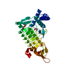
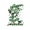
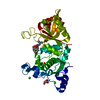
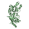
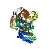
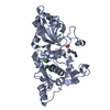
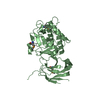
| Title | Crystal structure of mouse HPF1 | ||||||
 Components Components | Histone PARylation factor 1 | ||||||
 Keywords Keywords | NUCLEAR PROTEIN / cofactor / ADP-ribosylation / DNA damage response | ||||||
| Function / homology |  Function and homology information Function and homology informationprotein ADP-ribosyltransferase-substrate adaptor activity / poly-ADP-D-ribose binding / DNA repair-dependent chromatin remodeling / site of DNA damage / double-strand break repair / histone binding / chromatin binding / DNA damage response / chromatin / nucleus Similarity search - Function | ||||||
| Biological species |  | ||||||
| Method |  X-RAY DIFFRACTION / SYNCHROTRON / MOLECULAR REPLACEMENT / Resolution: 1.71 Å X-RAY DIFFRACTION / SYNCHROTRON / MOLECULAR REPLACEMENT / Resolution: 1.71 Å | ||||||
 Authors Authors | Sun, F.H. / Yun, C.H. | ||||||
| Funding support |  China, 1items China, 1items
| ||||||
 Citation Citation | Journal: Nat Commun / Year: 2021 Title: HPF1 remodels the active site of PARP1 to enable the serine ADP-ribosylation of histones. Authors: Sun, F.H. / Zhao, P. / Zhang, N. / Kong, L.L. / Wong, C.C.L. / Yun, C.H. | ||||||
| History |
|
- Structure visualization
| Structure viewer | Molecule: MolmilJmol/JSmol |
|---|
- Downloads & links
-Download
| PDBx/mmCIF format | 6m3h.cif.gz | 88.8 KB | Display | PDBx/mmCIF format |
|---|---|---|---|---|
| PDB format | pdb6m3h.ent.gz | 59 KB | Display | PDB format |
| PDBx/mmJSON format | 6m3h.json.gz | Tree view | PDBx/mmJSON format | |
| Others |  Other downloads Other downloads |
-Validation report
| Arichive directory | https://data.pdbj.org/pub/pdb/validation_reports/m3/6m3hftp://data.pdbj.org/pub/pdb/validation_reports/m3/6m3h | HTTPS FTP |
|---|
-Related structure data
| Related structure data | 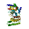 6m3gSC  6m3iC S: Starting model for refinement C: citing same article ( |
|---|---|
| Similar structure data |


-Links
 PDBj
PDBj- Assembly
| Deposited unit | 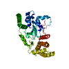
| ||||||||||||
|---|---|---|---|---|---|---|---|---|---|---|---|---|---|
| 1 |
| ||||||||||||
| Unit cell |
|
-Components
| #1: Protein | Mass: 39352.246 Da / Num. of mol.: 1 Source method: isolated from a genetically manipulated source Source: (gene. exp.) Production host: References: UniProt: Q8CFE2 |
|---|---|
| #2: Water | ChemComp-HOH /  Mass: 18.015 Da / Num. of mol.: 274 / Source method: isolated from a natural source / Formula: H2O Mass: 18.015 Da / Num. of mol.: 274 / Source method: isolated from a natural source / Formula: H2O |
-Experimental details
-Experiment
| Experiment | Method: X-RAY DIFFRACTION / Number of used crystals: 1 |
|---|
- Sample preparation
| Crystal | Density Matthews: 2.28 Å3/Da / Density % sol: 45.95 % |
|---|---|
| Crystal grow | Temperature: 300 K / Method: vapor diffusion Details: 0.1M Tris-pH8.3, 0.2 M Potassium citrate tribasic monohydrate, 20% w/v PEG 3350, |
-Data collection
| Diffraction | Mean temperature: 100 K / Serial crystal experiment: N |
|---|---|
| Diffraction source | Source: SYNCHROTRON / Site: SSRF / Beamline: BL18U1 / Wavelength: 0.97849 Å |
| Detector | Type: Nonius Kappa CCD / Detector: CCD / Date: Dec 7, 2018 |
| Radiation | Monochromator: Si(111) crystals / Protocol: SINGLE WAVELENGTH / Monochromatic (M) / Laue (L): M / Scattering type: x-ray |
| Radiation wavelength | Wavelength: 0.97849 Å / Relative weight: 1 |
| Reflection | Resolution: 1.71→50 Å / Num. obs: 39364 / % possible obs: 99.3 % / Redundancy: 5.7 % / Biso Wilson estimate: 21.62 Å2 / Rpim(I) all: 0.023 / Net I/σ(I): 31.1 |
| Reflection shell | Resolution: 1.71→1.77 Å / Num. unique obs: 2485 / Rpim(I) all: 0.351 |
- Processing
| Software |
| |||||||||||||||||||||||||||||||||||||||||||||||||||||||||||||||||||||||||||||||||||||||||||||||||||||||||
|---|---|---|---|---|---|---|---|---|---|---|---|---|---|---|---|---|---|---|---|---|---|---|---|---|---|---|---|---|---|---|---|---|---|---|---|---|---|---|---|---|---|---|---|---|---|---|---|---|---|---|---|---|---|---|---|---|---|---|---|---|---|---|---|---|---|---|---|---|---|---|---|---|---|---|---|---|---|---|---|---|---|---|---|---|---|---|---|---|---|---|---|---|---|---|---|---|---|---|---|---|---|---|---|---|---|---|
| Refinement | Method to determine structure: MOLECULAR REPLACEMENT Starting model: 6M3G Resolution: 1.71→44.36 Å / SU ML: 0.1648 / Cross valid method: NONE / σ(F): 1.35 / Phase error: 21.0794 / Stereochemistry target values: GeoStd + Monomer Library
| |||||||||||||||||||||||||||||||||||||||||||||||||||||||||||||||||||||||||||||||||||||||||||||||||||||||||
| Solvent computation | Shrinkage radii: 0.9 Å / VDW probe radii: 1.11 Å / Solvent model: FLAT BULK SOLVENT MODEL | |||||||||||||||||||||||||||||||||||||||||||||||||||||||||||||||||||||||||||||||||||||||||||||||||||||||||
| Displacement parameters | Biso mean: 25.28 Å2 | |||||||||||||||||||||||||||||||||||||||||||||||||||||||||||||||||||||||||||||||||||||||||||||||||||||||||
| Refinement step | Cycle: LAST / Resolution: 1.71→44.36 Å
| |||||||||||||||||||||||||||||||||||||||||||||||||||||||||||||||||||||||||||||||||||||||||||||||||||||||||
| Refine LS restraints |
| |||||||||||||||||||||||||||||||||||||||||||||||||||||||||||||||||||||||||||||||||||||||||||||||||||||||||
| LS refinement shell |
|
