 Movie
Movie Controller

[English] 日本語
 Yorodumi
- PDB-1tl9: High resolution crystal structure of calpain I protease core in c... -
+ Open data

- Basic information
| Entry | Database: PDB / ID: 1tl9 | ||||||
|---|---|---|---|---|---|---|---|
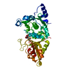
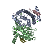
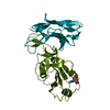
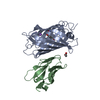
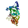
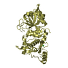
| Title | High resolution crystal structure of calpain I protease core in complex with leupeptin | ||||||
 Components Components |
| ||||||
 Keywords Keywords | HYDROLASE/HYDROLASE INHIBITOR / Covalently-linked inhibitor at the active site cysteine forms a hemithioacetal / hydrolase / HYDROLASE-HYDROLASE INHIBITOR COMPLEX | ||||||
| Function / homology |  Function and homology information Function and homology informationcalpain-1 / Degradation of the extracellular matrix / regulation of catalytic activity / protein catabolic process at postsynapse / positive regulation of leukocyte tethering or rolling / mammary gland involution / calcium-dependent cysteine-type endopeptidase activity / negative regulation of actin filament polymerization / self proteolysis / cornified envelope ...calpain-1 / Degradation of the extracellular matrix / regulation of catalytic activity / protein catabolic process at postsynapse / positive regulation of leukocyte tethering or rolling / mammary gland involution / calcium-dependent cysteine-type endopeptidase activity / negative regulation of actin filament polymerization / self proteolysis / cornified envelope / receptor catabolic process / response to angiotensin / positive regulation of vascular permeability / response to arsenic-containing substance / negative regulation of non-canonical NF-kappaB signal transduction / Neutrophil degranulation / protein autoprocessing / cytoskeletal protein binding / positive regulation of cardiac muscle cell apoptotic process / protein catabolic process / cellular response to hydrogen peroxide / peptidase activity / presynapse / lysosome / postsynaptic density / postsynapse / calcium ion binding / glutamatergic synapse / enzyme binding / mitochondrion / proteolysis / membrane / plasma membrane / cytoplasm / cytosol Similarity search - Function | ||||||
| Biological species |   Actinomycetes Streptomyces roseus MA 839-A1 (bacteria) Actinomycetes Streptomyces roseus MA 839-A1 (bacteria) | ||||||
| Method |  X-RAY DIFFRACTION / MOLECULAR REPLACEMENT / Resolution: 1.8 Å X-RAY DIFFRACTION / MOLECULAR REPLACEMENT / Resolution: 1.8 Å | ||||||
 Authors Authors | Moldoveanu, T. / Campbell, R.L. / Cuerrier, D. / Davies, P.L. | ||||||
 Citation Citation | Journal: J.Mol.Biol. / Year: 2004 Title: Crystal Structures of Calpain-E64 and -Leupeptin Inhibitor Complexes Reveal Mobile Loops Gating the Active Site Authors: Moldoveanu, T. / Campbell, R.L. / Cuerrier, D. / Davies, P.L. #1: Journal: Cell(Cambridge,Mass.) / Year: 2002Title: A calcium switch aligns the active site of calpain Authors: Moldoveanu, T. / Hosfield, C.M. / Lim, D. / Elce, J.S. / Jia, Z. / Davies, P.L. #2: Journal: Nat.Struct.Mol.Biol. / Year: 2003Title: calpain silencing by a reversible intrinsic mechanism Authors: Moldoveanu, T. / Hosfield, C.M. / Lim, D. / Jia, Z. / Davies, P.L. | ||||||
| History |
|
- Structure visualization
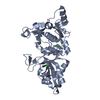
| Structure viewer | Molecule: MolmilJmol/JSmol |
|---|
- Downloads & links
-Download
| PDBx/mmCIF format | 1tl9.cif.gz | 87.9 KB | Display | PDBx/mmCIF format |
|---|---|---|---|---|
| PDB format | pdb1tl9.ent.gz | 64.1 KB | Display | PDB format |
| PDBx/mmJSON format | 1tl9.json.gz | Tree view | PDBx/mmJSON format | |
| Others |  Other downloads Other downloads |
-Validation report
| Arichive directory | https://data.pdbj.org/pub/pdb/validation_reports/tl/1tl9ftp://data.pdbj.org/pub/pdb/validation_reports/tl/1tl9 | HTTPS FTP |
|---|
-Related structure data


-Links
 PDBj
PDBj


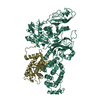
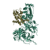
- Assembly
| Deposited unit | 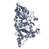
| ||||||||
|---|---|---|---|---|---|---|---|---|---|
| 1 |
| ||||||||
| Unit cell |
|
-Components
| #1: Protein | Mass: 38804.551 Da / Num. of mol.: 1 / Fragment: residues 27-356 Source method: isolated from a genetically manipulated source Source: (gene. exp.) | ||||||||
|---|---|---|---|---|---|---|---|---|---|
| #2: Protein/peptide | 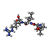  Type: Oligopeptide / Class: Enzyme inhibitor / Mass: 429.578 Da / Num. of mol.: 1 / Source method: obtained synthetically Type: Oligopeptide / Class: Enzyme inhibitor / Mass: 429.578 Da / Num. of mol.: 1 / Source method: obtained syntheticallySource: (synth.) Actinomycetes Streptomyces roseus MA 839-A1 (bacteria)References: NOR: NOR00487, LEUPEPTIN | ||||||||
| #3: Chemical |   Mass: 40.078 Da / Num. of mol.: 2 / Source method: obtained synthetically / Formula: Ca Mass: 40.078 Da / Num. of mol.: 2 / Source method: obtained synthetically / Formula: Ca#4: Water | ChemComp-HOH / |  Mass: 18.015 Da / Num. of mol.: 287 / Source method: isolated from a natural source / Formula: H2O Mass: 18.015 Da / Num. of mol.: 287 / Source method: isolated from a natural source / Formula: H2OCompound details | THE LEUPEPTIN IS COVALENTLY | Has protein modification | Y | Sequence details | THERE IS A DISCREPANCY IN THE NORINE AND PDB NUMBERING, AS NORINE COUNTS ACE AND LEU TOGETHER AS ...THERE IS A DISCREPANC | |
-Experimental details
-Experiment
| Experiment | Method: X-RAY DIFFRACTION / Number of used crystals: 1 |
|---|
- Sample preparation
| Crystal | Density Matthews: 2.2 Å3/Da / Density % sol: 44.17 % |
|---|---|
| Crystal grow | pH: 6 Details: sodium chloride, calcium chloride, MES, pH 6.0, VAPOR DIFFUSION, HANGING DROP, pH 6.00 |
-Data collection
| Diffraction | Mean temperature: 100 K |
|---|---|
| Diffraction source | Source: ROTATING ANODE / Type: RIGAKU RU200 / Wavelength: 1.5418 |
| Detector | Type: MARRESEARCH / Detector: IMAGE PLATE / Date: Apr 2, 2004 / Details: OSMIC MIRRORS |
| Radiation | Monochromator: OSMIC MIRRORS / Protocol: SINGLE WAVELENGTH / Monochromatic (M) / Laue (L): M / Scattering type: x-ray |
| Radiation wavelength | Wavelength: 1.5418 Å / Relative weight: 1 |
| Reflection | Resolution: 1.8→50 Å / Num. obs: 31962 / % possible obs: 95 % / Observed criterion σ(I): 2 / Rmerge(I) obs: 0.069 / Rsym value: 0.072 |
| Reflection shell | Resolution: 1.8→1.86 Å / Rmerge(I) obs: 0.147 / Mean I/σ(I) obs: 14.9 / Num. measured obs: 3221 / Rsym value: 0.152 |
- Processing
| Software |
| ||||||||||||||||||||||||||||||||||||||||||||||||||||||||||||
|---|---|---|---|---|---|---|---|---|---|---|---|---|---|---|---|---|---|---|---|---|---|---|---|---|---|---|---|---|---|---|---|---|---|---|---|---|---|---|---|---|---|---|---|---|---|---|---|---|---|---|---|---|---|---|---|---|---|---|---|---|---|
| Refinement | Method to determine structure: MOLECULAR REPLACEMENT Starting model: PDB ENTRY 1KXRB Resolution: 1.8→50 Å / Cross valid method: THROUGHOUT / σ(F): 2 / Stereochemistry target values: ENGH & HUBER
| ||||||||||||||||||||||||||||||||||||||||||||||||||||||||||||
| Refinement step | Cycle: LAST / Resolution: 1.8→50 Å
| ||||||||||||||||||||||||||||||||||||||||||||||||||||||||||||
| Refine LS restraints |
|
