[English] 日本語
Yorodumi
- PDB-6bgp: Crystal Structure of Human Calpain-3 Protease Core Mutant-C129A -

+
Open data


ID or keywords:

Loading...

-
Basic information

Entry
Database: PDB / ID: 6bgp
TitleCrystal Structure of Human Calpain-3 Protease Core Mutant-C129A
ComponentsCalpain-3
KeywordsHYDROLASE / Calcium binding / Cysteine protease / Calpain / P94
Function / homology
Function and homology information


calpain-3 / positive regulation of satellite cell activation involved in skeletal muscle regeneration / ligase regulator activity / calcium-dependent self proteolysis / G1 to G0 transition involved in cell differentiation / regulation of myoblast differentiation / cellular response to salt stress / myofibril assembly / muscle structure development / calcium-dependent cysteine-type endopeptidase activity ...calpain-3 / positive regulation of satellite cell activation involved in skeletal muscle regeneration / ligase regulator activity / calcium-dependent self proteolysis / G1 to G0 transition involved in cell differentiation / regulation of myoblast differentiation / cellular response to salt stress / myofibril assembly / muscle structure development / calcium-dependent cysteine-type endopeptidase activity / sodium ion binding / self proteolysis / regulation of catalytic activity / negative regulation of protein sumoylation / protein localization to membrane / regulation of canonical NF-kappaB signal transduction / response to muscle activity / muscle organ development / structural constituent of muscle / muscle cell cellular homeostasis / myofibril / sarcomere organization / positive regulation of proteolysis / catalytic activity / cysteine-type peptidase activity / titin binding / T-tubule / cellular response to calcium ion / Degradation of the extracellular matrix / positive regulation of release of sequestered calcium ion into cytosol / protein destabilization / protein catabolic process / Z disc / response to calcium ion / peptidase activity / positive regulation of NF-kappaB transcription factor activity / protein-containing complex assembly / molecular adaptor activity / negative regulation of DNA-templated transcription / apoptotic process / calcium ion binding / nucleolus / negative regulation of apoptotic process / positive regulation of DNA-templated transcription / protein-containing complex / proteolysis / identical protein binding / nucleus / plasma membrane / cytoplasm / cytosol
Similarity search - Function
Calpain-3 / Unstructured region on Calpain-3 / Calpain subdomain III / Peptidase C2, calpain, domain III / calpain_III / Peptidase C2, calpain, large subunit, domain III / Peptidase C2, calpain family / Calpain large subunit, domain III superfamily / Calpain large subunit, domain III / Peptidase C2, calpain, catalytic domain ...Calpain-3 / Unstructured region on Calpain-3 / Calpain subdomain III / Peptidase C2, calpain, domain III / calpain_III / Peptidase C2, calpain, large subunit, domain III / Peptidase C2, calpain family / Calpain large subunit, domain III superfamily / Calpain large subunit, domain III / Peptidase C2, calpain, catalytic domain / Calpain family cysteine protease / Cysteine proteinase, calpain-type, catalytic domain profile. / Calpain-like thiol protease family. / EF-hand domain / EF-hand domain pair / Cysteine proteinases / Cysteine peptidase, cysteine active site / Eukaryotic thiol (cysteine) proteases cysteine active site. / Cathepsin B; Chain A / Papain-like cysteine peptidase superfamily / EF-hand, calcium binding motif / EF-Hand 1, calcium-binding site / EF-hand calcium-binding domain. / EF-hand calcium-binding domain profile. / EF-hand domain / EF-hand domain pair / Alpha-Beta Complex / Alpha Beta
Similarity search - Domain/homology
Biological speciesHomo sapiens (human)
MethodX-RAY DIFFRACTION / SYNCHROTRON / MOLECULAR REPLACEMENT / Resolution: 2.75 Å
AuthorsYe, Q. / Campbell, R.L. / Davies, P.L.
Funding support Canada, 1items
OrganizationGrant numberCountry
Canadian Institutes of Health Research (CIHR)MOP 74681 Canada
CitationJournal: J. Biol. Chem. / Year: 2018
Title: Structures of human calpain-3 protease core with and without bound inhibitor reveal mechanisms of calpain activation.
Authors: Ye, Q. / Campbell, R.L. / Davies, P.L.
History
DepositionOct 29, 2017Deposition site: RCSB / Processing site: RCSB
Revision 1.0Feb 7, 2018Provider: repository / Type: Initial release
Revision 1.1Feb 14, 2018Group: Database references / Category: citation
Item: _citation.country / _citation.journal_abbrev ..._citation.country / _citation.journal_abbrev / _citation.journal_id_ASTM / _citation.journal_id_CSD / _citation.journal_id_ISSN / _citation.pdbx_database_id_DOI / _citation.pdbx_database_id_PubMed / _citation.title
Revision 1.2May 2, 2018Group: Data collection / Database references / Category: citation
Item: _citation.journal_volume / _citation.page_first / _citation.page_last
Revision 1.3Jan 8, 2020Group: Author supporting evidence / Category: pdbx_audit_support / Item: _pdbx_audit_support.funding_organization
Revision 1.4Oct 4, 2023Group: Data collection / Database references / Refinement description
Category: chem_comp_atom / chem_comp_bond ...chem_comp_atom / chem_comp_bond / database_2 / pdbx_initial_refinement_model / struct_ncs_dom_lim
Item: _database_2.pdbx_DOI / _database_2.pdbx_database_accession ..._database_2.pdbx_DOI / _database_2.pdbx_database_accession / _struct_ncs_dom_lim.beg_auth_comp_id / _struct_ncs_dom_lim.beg_label_asym_id / _struct_ncs_dom_lim.beg_label_comp_id / _struct_ncs_dom_lim.beg_label_seq_id / _struct_ncs_dom_lim.end_auth_comp_id / _struct_ncs_dom_lim.end_label_asym_id / _struct_ncs_dom_lim.end_label_comp_id / _struct_ncs_dom_lim.end_label_seq_id

-
Structure visualization

Structure viewerMolecule:
MolmilJmol/JSmol

Downloads & links

-
Assembly

Deposited unit
A: Calpain-3
B: Calpain-3
C: Calpain-3
D: Calpain-3
hetero molecules


Theoretical massNumber of molelcules
Total (without water)178,15916
Polymers177,6964
Non-polymers46212
Water90150
1
A: Calpain-3
hetero molecules


Theoretical massNumber of molelcules
Total (without water)44,5404
Polymers44,4241
Non-polymers1163
Water181
TypeNameSymmetry operationNumber
identity operation1_555x,y,z1
2
B: Calpain-3
hetero molecules


Theoretical massNumber of molelcules
Total (without water)44,5404
Polymers44,4241
Non-polymers1163
Water181
TypeNameSymmetry operationNumber
identity operation1_555x,y,z1
3
C: Calpain-3
hetero molecules


Theoretical massNumber of molelcules
Total (without water)44,5404
Polymers44,4241
Non-polymers1163
Water181
TypeNameSymmetry operationNumber
identity operation1_555x,y,z1
4
D: Calpain-3
hetero molecules


Theoretical massNumber of molelcules
Total (without water)44,5404
Polymers44,4241
Non-polymers1163
Water181
TypeNameSymmetry operationNumber
identity operation1_555x,y,z1
Unit cell
Length a, b, c (Å)60.100, 106.180, 225.620
Angle α, β, γ (deg.)90.000, 90.000, 90.000
Int Tables number19
Space group name H-MP212121
Noncrystallographic symmetry (NCS)NCS domain:
IDEns-IDDetails
11A
21B
12A
22C
13A
23D
14B
24C
15B
25D
16C
26D

NCS domain segments:

Component-ID: _ / Beg auth comp-ID: GLU / Beg label comp-ID: GLU / Refine code: _

Dom-IDEns-IDEnd auth comp-IDEnd label comp-IDAuth asym-IDLabel asym-IDAuth seq-IDLabel seq-ID
11THRTHRAA58 - 41713 - 372
21THRTHRBB58 - 41713 - 372
12THRTHRAA58 - 41713 - 372
22THRTHRCC58 - 41713 - 372
13THRTHRAA58 - 41713 - 372
23THRTHRDD58 - 41713 - 372
14THRTHRBB58 - 41713 - 372
24THRTHRCC58 - 41713 - 372
15ASPASPBB58 - 41913 - 374
25ASPASPDD58 - 41913 - 374
16THRTHRCC58 - 41713 - 372
26THRTHRDD58 - 41713 - 372

NCS ensembles :
ID
1
2
3
4
5
6

-
Components

#1: Protein
Calpain-3 / Calcium-activated neutral proteinase 3 / CANP 3 / Calpain L3 / Calpain p94 / Muscle-specific ...Calcium-activated neutral proteinase 3 / CANP 3 / Calpain L3 / Calpain p94 / Muscle-specific calcium-activated neutral protease 3 / New calpain 1 / nCL-1


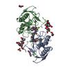
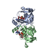
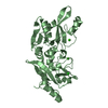
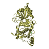
Mass: 44424.109 Da / Num. of mol.: 4 / Mutation: C129A
Source method: isolated from a genetically manipulated source
Details: Muscle-specific calcium-activated neutral protease 3 that is activated by autoproteolytic cleavage of insertion sequence 1 (IS1), which allows substrates and inhibitors gain access to the active site.
Source: (gene. exp.) Homo sapiens (human) / Gene: CAPN3, CANP3, CANPL3, NCL1 / Plasmid: pET24a / Production host: Escherichia coli (E. coli) / Strain (production host): BL21 (DE3) / References: UniProt: P20807, calpain-3
#2: Chemical
ChemComp-CA / CALCIUM ION


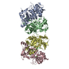
Mass: 40.078 Da / Num. of mol.: 8 / Source method: obtained synthetically / Formula: Ca
#3: Chemical
ChemComp-CL / CHLORIDE ION


Mass: 35.453 Da / Num. of mol.: 4 / Source method: obtained synthetically / Formula: Cl
#4: Water ChemComp-HOH / water


Mass: 18.015 Da / Num. of mol.: 50 / Source method: isolated from a natural source / Formula: H2O

-
Experimental details

-
Experiment

ExperimentMethod: X-RAY DIFFRACTION / Number of used crystals: 1

-
Sample preparation

CrystalDensity Matthews: 2.03 Å3/Da / Density % sol: 39.28 %
Crystal growTemperature: 295.15 K / Method: vapor diffusion, hanging drop / pH: 6.5 / Details: PEG 8000, MES, calcium chloride

-
Data collection

DiffractionMean temperature: 100 K
Diffraction sourceSource: SYNCHROTRON / Site: NSLS / Beamline: X6A / Wavelength: 1.0781 Å
DetectorType: ADSC QUANTUM 210 / Detector: CCD / Date: Sep 18, 2014 / Details: Oxford Danfysik toroidal focusing mirror
RadiationMonochromator: channel cut monochromator, Si(111) / Protocol: SINGLE WAVELENGTH / Monochromatic (M) / Laue (L): M / Scattering type: x-ray
Radiation wavelengthWavelength: 1.0781 Å / Relative weight: 1
ReflectionResolution: 2.75→30 Å / Num. obs: 38363 / % possible obs: 99.7 % / Redundancy: 14.69 % / CC1/2: 0.998 / Rrim(I) all: 0.17 / Χ2: 0.96 / Net I/σ(I): 16.42
Reflection shellResolution: 2.75→2.77 Å / Redundancy: 14.97 % / Mean I/σ(I) obs: 2.38 / Num. unique obs: 8698 / CC1/2: 0.76 / Rrim(I) all: 1.44 / Χ2: 0.93 / % possible all: 100

-
Processing

Software
NameVersionClassification
REFMAC5.8.0073refinement
PDB_EXTRACT3.22data extraction
XDSdata reduction
XSCALEdata scaling
PHASERphasing
RefinementMethod to determine structure: MOLECULAR REPLACEMENT
Starting model: 6BDT
Resolution: 2.75→17.94 Å / Cor.coef. Fo:Fc: 0.939 / Cor.coef. Fo:Fc free: 0.906 / SU B: 16.746 / SU ML: 0.325 / Cross valid method: THROUGHOUT / σ(F): 0 / ESU R Free: 0.385
Details: HYDROGENS HAVE BEEN ADDED IN THE RIDING POSITIONS U VALUES : REFINED INDIVIDUALLY
RfactorNum. reflection% reflectionSelection details
Rfree0.2584 1916 5 %RANDOM
Rwork0.2092 ---
obs0.2117 36402 99.54 %-
Solvent computationIon probe radii: 0.8 Å / Shrinkage radii: 0.8 Å / VDW probe radii: 1.2 Å
Displacement parametersBiso max: 164.19 Å2 / Biso mean: 68.376 Å2 / Biso min: 20 Å2
Baniso -1Baniso -2Baniso -3
1-1.82 Å20 Å20 Å2
2--0.33 Å2-0 Å2
3----2.14 Å2
Refinement stepCycle: final / Resolution: 2.75→17.94 Å
ProteinNucleic acidLigandSolventTotal
Num. atoms10254 0 12 50 10316
Biso mean--50.31 51.17 -
Num. residues----1244
Refine LS restraints
Refine-IDTypeDev idealDev ideal targetNumber
X-RAY DIFFRACTIONr_bond_refined_d0.0080.01910550
X-RAY DIFFRACTIONr_bond_other_d0.0040.029609
X-RAY DIFFRACTIONr_angle_refined_deg1.2041.92614274
X-RAY DIFFRACTIONr_angle_other_deg1.066322139
X-RAY DIFFRACTIONr_dihedral_angle_1_deg5.64551235
X-RAY DIFFRACTIONr_dihedral_angle_2_deg34.67324.158558
X-RAY DIFFRACTIONr_dihedral_angle_3_deg14.224151774
X-RAY DIFFRACTIONr_dihedral_angle_4_deg13.1181556
X-RAY DIFFRACTIONr_chiral_restr0.0720.21453
X-RAY DIFFRACTIONr_gen_planes_refined0.0060.0211953
X-RAY DIFFRACTIONr_gen_planes_other0.0040.022631
Refine LS restraints NCS

Refine-ID: X-RAY DIFFRACTION / Type: interatomic distance / Weight position: 0.05

Ens-IDDom-IDAuth asym-IDNumberRms dev position (Å)
11A177910.11
12B177910.11
21A175600.11
22C175600.11
31A177070.12
32D177070.12
41B182870.11
42C182870.11
51B186210.11
52D186210.11
61C182730.11
62D182730.11
LS refinement shellResolution: 2.75→2.82 Å / Rfactor Rfree error: 0 / Total num. of bins used: 20
RfactorNum. reflection% reflection
Rfree0.356 136 -
Rwork0.312 2597 -
all-2733 -
obs--100 %

+
About Yorodumi

-
News

-
Feb 9, 2022. New format data for meta-information of EMDB entries

New format data for meta-information of EMDB entries

  • Version 3 of the EMDB header file is now the official format.
  • The previous official version 1.9 will be removed from the archive.

Related info.:EMDB header

External links:wwPDB to switch to version 3 of the EMDB data model

-
Aug 12, 2020. Covid-19 info

Covid-19 info

URL: https://pdbjlvh1.pdbj.org/emnavi/covid19.php

New page: Covid-19 featured information page in EM Navigator.

Related info.:Covid-19 info / Mar 5, 2020. Novel coronavirus structure data

+
Mar 5, 2020. Novel coronavirus structure data

Novel coronavirus structure data

Related info.:Yorodumi Speices / Aug 12, 2020. Covid-19 info

External links:COVID-19 featured content - PDBj / Molecule of the Month (242):Coronavirus Proteases

+
Jan 31, 2019. EMDB accession codes are about to change! (news from PDBe EMDB page)

EMDB accession codes are about to change! (news from PDBe EMDB page)

  • The allocation of 4 digits for EMDB accession codes will soon come to an end. Whilst these codes will remain in use, new EMDB accession codes will include an additional digit and will expand incrementally as the available range of codes is exhausted. The current 4-digit format prefixed with “EMD-” (i.e. EMD-XXXX) will advance to a 5-digit format (i.e. EMD-XXXXX), and so on. It is currently estimated that the 4-digit codes will be depleted around Spring 2019, at which point the 5-digit format will come into force.
  • The EM Navigator/Yorodumi systems omit the EMD- prefix.

Related info.:Q: What is EMD? / ID/Accession-code notation in Yorodumi/EM Navigator

External links:EMDB Accession Codes are Changing Soon! / Contact to PDBj

+
Jul 12, 2017. Major update of PDB

Major update of PDB

  • wwPDB released updated PDB data conforming to the new PDBx/mmCIF dictionary.
  • This is a major update changing the version number from 4 to 5, and with Remediation, in which all the entries are updated.
  • In this update, many items about electron microscopy experimental information are reorganized (e.g. em_software).
  • Now, EM Navigator and Yorodumi are based on the updated data.

External links:wwPDB Remediation / Enriched Model Files Conforming to OneDep Data Standards Now Available in the PDB FTP Archive

-
Yorodumi

Thousand views of thousand structures

  • Yorodumi is a browser for structure data from EMDB, PDB, SASBDB, etc.
  • This page is also the successor to EM Navigator detail page, and also detail information page/front-end page for Omokage search.
  • The word "yorodu" (or yorozu) is an old Japanese word meaning "ten thousand". "mi" (miru) is to see.

Related info.:EMDB / PDB / SASBDB / Comparison of 3 databanks / Yorodumi Search / Aug 31, 2016. New EM Navigator & Yorodumi / Yorodumi Papers / Jmol/JSmol / Function and homology information / Changes in new EM Navigator and Yorodumi

Read more