 Movie
Movie Controller


+ Open data

- Basic information
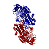
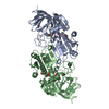
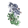
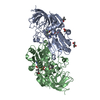
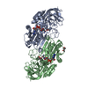
| Entry | Database: PDB / ID: 1ye3 | ||||||
|---|---|---|---|---|---|---|---|
| Title | HORSE LIVER ALCOHOL DEHYDROGENASE APOENZYME | ||||||
 Components Components | Alcohol dehydrogenase E chain | ||||||
 Keywords Keywords | OXIDOREDUCTASE / DEHYDROGENASE / ALCOHOL / LIVER / APOENZYME / METHYLPENTANEDIOL | ||||||
| Function / homology |  Function and homology information Function and homology informationall-trans-retinol dehydrogenase (NAD+) activity / alcohol dehydrogenase / retinoic acid metabolic process / retinol metabolic process / zinc ion binding / cytosol Similarity search - Function | ||||||
| Biological species |  | ||||||
| Method |  X-RAY DIFFRACTION / SYNCHROTRON / MOLECULAR REPLACEMENT / Resolution: 1.59 Å X-RAY DIFFRACTION / SYNCHROTRON / MOLECULAR REPLACEMENT / Resolution: 1.59 Å | ||||||
 Authors Authors | Plapp, B.V. / Savarimuthu, B.R. / Ramaswamy, S. | ||||||
 Citation Citation | Journal: To be Published Title: Horse Liver Alcohol Dehydrogenase Apoenzyme Authors: Plapp, B.V. / Savarimuthu, B.R. / Ramaswamy, S. #1: Journal: J.Mol.Biol. / Year: 1976Title: Three-Dimensional Structure of Horse Liver Alcohol Dehydrogenase at 2.4 Angstroms Resolution Authors: Eklund, H. / Nordstrom, B. / Zeppezauer, E. / Soderlund, G. / Ohlsson, I. / Boiwe, T. / Soderberg, B.-O. / Tapia, O. / Branden, C.-I. / Akeson, A. #2: Journal: J.Mol.Biol. / Year: 1981Title: Structure of a Triclinic Ternary Complex of Horse Liver Alcohol Dehydrogenase at 2.9 Angstroms Resolution Authors: Eklund, H. / Samama, J.-P. / Wallen, L. / Branden, C.-I. / Akeson, A. / A Jones, T. #3: Journal: Biochemistry / Year: 1999Title: Substitutions in the Flexible Loop of Horse Liver Alcohol Dehydrogenase Hinder the Conformational Change and Unmask Hydrogen Transfer Authors: Ramaswamy, S. / Park, D.H. / Plapp, B.V. #4: Journal: Biochemistry / Year: 2001Title: Contributions of Valine-292 in the Nicotinamide Binding Site of Liver Alcohol Dehydrogenase and Dynamics to Catalysis Authors: Rubach, J.K. / Ramaswamy, S. / Plapp, B.V. #5: Journal: J.Biol.Chem. / Year: 1982Title: Binding of Substrate in a Ternary Complex of Horse Liver Alcohol Dehydrogenase Authors: Eklund, H. / Plapp, B.V. / Samama, J.-P. / Branden, C.-I. #6: Journal: Biochemistry / Year: 1994Title: Structures of Horse Liver Alcohol Dehydrogenase Complexed with Nad+ and Substituted Benzyl Alcohols Authors: Ramaswamy, S. / Eklund, H. / Plapp, B.V. #7: Journal: Biochemistry / Year: 2002Title: Mobility of Fluorobenzyl Alcohols Bound to Liver Alcohol Dehydrogenases as Determined by NMR and X-Ray Crystallographic Studies Authors: Rubach, J.K. / Plapp, B.V. #8: Journal: Biochemistry / Year: 2003Title: Amino Acid Residues in the Nicotinamide Binding Site Contribute to Catalysis by Horse Liver Alcohol Dehydrogenase Authors: Rubach, J.K. / Plapp, B.V. #9: Journal: Biochemistry / Year: 1984Title: Crystallographic Investigations of Nicotinamide Adenine Dinucleotide Binding to Horse Liver Alcohol Dehydrogenase Authors: Eklund, H. / Samama, J.-P. / Jones, T.A. #10: Journal: Biochemistry / Year: 1982Title: Crystal Structure Determinations of Coenzyme Analogue and Substrate Complexes of Liver Alcohol Dehydrogenase. Binding of 1,4,5,6-Tetrahydronicotinamide Adenine Dinucleotide and Trans-4-(N,N- ...Title: Crystal Structure Determinations of Coenzyme Analogue and Substrate Complexes of Liver Alcohol Dehydrogenase. Binding of 1,4,5,6-Tetrahydronicotinamide Adenine Dinucleotide and Trans-4-(N,N-Dimethylamino)Cinnamaldehyde to the Enzyme Authors: Cedergren-Zeppezauer, E. / Samama, J.-P. / Eklund, H. | ||||||
| History |
|
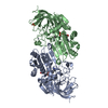
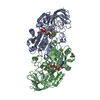
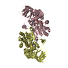
- Structure visualization
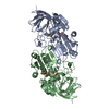
| Structure viewer | Molecule: MolmilJmol/JSmol |
|---|
- Downloads & links
-Download
| PDBx/mmCIF format | 1ye3.cif.gz | 153.7 KB | Display | PDBx/mmCIF format |
|---|---|---|---|---|
| PDB format | pdb1ye3.ent.gz | 120 KB | Display | PDB format |
| PDBx/mmJSON format | 1ye3.json.gz | Tree view | PDBx/mmJSON format | |
| Others |  Other downloads Other downloads |
-Validation report
| Arichive directory | https://data.pdbj.org/pub/pdb/validation_reports/ye/1ye3ftp://data.pdbj.org/pub/pdb/validation_reports/ye/1ye3 | HTTPS FTP |
|---|
-Related structure data
| Related structure data | 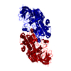 8adhS S: Starting model for refinement |
|---|---|
| Similar structure data |

-Links
 PDBj
PDBj

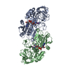
- Assembly
| Deposited unit | 
| ||||||||
|---|---|---|---|---|---|---|---|---|---|
| 1 | 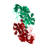
| ||||||||
| Unit cell |
|
-Components
| #1: Protein | Mass: 39853.273 Da / Num. of mol.: 1 / Source method: isolated from a natural source / Details: liver / Source: (natural) | ||||
|---|---|---|---|---|---|
| #2: Chemical |   Mass: 65.409 Da / Num. of mol.: 2 / Source method: obtained synthetically / Formula: Zn Mass: 65.409 Da / Num. of mol.: 2 / Source method: obtained synthetically / Formula: Zn#3: Chemical | ChemComp-MPD / ( |   Mass: 118.174 Da / Num. of mol.: 1 / Source method: obtained synthetically / Formula: C6H14O2 / Comment: precipitant*YM Mass: 118.174 Da / Num. of mol.: 1 / Source method: obtained synthetically / Formula: C6H14O2 / Comment: precipitant*YM#4: Water | ChemComp-HOH / |  Mass: 18.015 Da / Num. of mol.: 200 / Source method: isolated from a natural source / Formula: H2O Mass: 18.015 Da / Num. of mol.: 200 / Source method: isolated from a natural source / Formula: H2O |
-Experimental details
-Experiment
| Experiment | Method: X-RAY DIFFRACTION / Number of used crystals: 1 |
|---|
- Sample preparation
| Crystal | Density Matthews: 2.36 Å3/Da / Density % sol: 47.79 % |
|---|---|
| Crystal grow | Temperature: 278 K / Method: microdialysis / pH: 8.4 Details: 50 mM Tris-HCl, 25% methylpentanediol, pH 8.4, MICRODIALYSIS, temperature 278K |
-Data collection
| Diffraction | Mean temperature: 100 K |
|---|---|
| Diffraction source | Source: SYNCHROTRON / Site: APS  / Beamline: 17-ID / Wavelength: 1 Å / Beamline: 17-ID / Wavelength: 1 Å |
| Detector | Type: ADSC QUANTUM 4 / Detector: CCD / Date: Jul 23, 2004 |
| Radiation | Protocol: SINGLE WAVELENGTH / Monochromatic (M) / Laue (L): M / Scattering type: x-ray |
| Radiation wavelength | Wavelength: 1 Å / Relative weight: 1 |
| Reflection | Resolution: 1.59→20.2 Å / Num. obs: 36732 / % possible obs: 71.9 % / Observed criterion σ(F): 1 / Observed criterion σ(I): 1 / Redundancy: 5.66 % / Biso Wilson estimate: 25.468 Å2 / Rmerge(I) obs: 0.075 / Net I/σ(I): 11.5 |
| Reflection shell | Resolution: 1.59→1.65 Å / Redundancy: 5.74 % / Rmerge(I) obs: 0.211 / Mean I/σ(I) obs: 4.7 / Num. unique all: 2918 / % possible all: 58 |
- Processing
| Software |
| ||||||||||||||||||||||||||||||||||||||||||||||||||||||||||||||||||||||||||||||||||||||||||||||||||||||||||||||
|---|---|---|---|---|---|---|---|---|---|---|---|---|---|---|---|---|---|---|---|---|---|---|---|---|---|---|---|---|---|---|---|---|---|---|---|---|---|---|---|---|---|---|---|---|---|---|---|---|---|---|---|---|---|---|---|---|---|---|---|---|---|---|---|---|---|---|---|---|---|---|---|---|---|---|---|---|---|---|---|---|---|---|---|---|---|---|---|---|---|---|---|---|---|---|---|---|---|---|---|---|---|---|---|---|---|---|---|---|---|---|---|
| Refinement | Method to determine structure: MOLECULAR REPLACEMENT Starting model: 8ADH Resolution: 1.59→20.2 Å / Cor.coef. Fo:Fc: 0.948 / Cor.coef. Fo:Fc free: 0.92 / SU B: 3.926 / SU ML: 0.069 / Isotropic thermal model: anisotropic / Cross valid method: THROUGHOUT / σ(F): 1 / ESU R: 0.2 / ESU R Free: 0.133 / Stereochemistry target values: MAXIMUM LIKELIHOOD Details: Positions for residues A269-A270 were not well-defined by the electron density. Several residues, including A306, have alternative conformations, but these were not modeled.
| ||||||||||||||||||||||||||||||||||||||||||||||||||||||||||||||||||||||||||||||||||||||||||||||||||||||||||||||
| Solvent computation | Ion probe radii: 0.8 Å / Shrinkage radii: 0.8 Å / VDW probe radii: 1.2 Å / Solvent model: BABINET MODEL WITH MASK | ||||||||||||||||||||||||||||||||||||||||||||||||||||||||||||||||||||||||||||||||||||||||||||||||||||||||||||||
| Displacement parameters | Biso mean: 35.47 Å2
| ||||||||||||||||||||||||||||||||||||||||||||||||||||||||||||||||||||||||||||||||||||||||||||||||||||||||||||||
| Refinement step | Cycle: LAST / Resolution: 1.59→20.2 Å
| ||||||||||||||||||||||||||||||||||||||||||||||||||||||||||||||||||||||||||||||||||||||||||||||||||||||||||||||
| Refine LS restraints |
| ||||||||||||||||||||||||||||||||||||||||||||||||||||||||||||||||||||||||||||||||||||||||||||||||||||||||||||||
| LS refinement shell | Resolution: 1.59→1.631 Å / Total num. of bins used: 20
|
