 Movie
Movie Controller

[English] 日本語
 Yorodumi
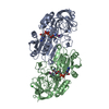
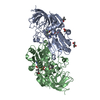
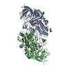
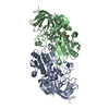
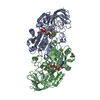
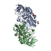
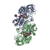
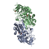
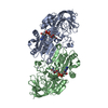
- PDB-1htb: CRYSTALLIZATION OF HUMAN BETA3 ALCOHOL DEHYDROGENASE (10 MG/ML) I... -
+ Open data

- Basic information
| Entry | Database: PDB / ID: 1htb | ||||||
|---|---|---|---|---|---|---|---|
| Title | CRYSTALLIZATION OF HUMAN BETA3 ALCOHOL DEHYDROGENASE (10 MG/ML) IN 100 MM SODIUM PHOSPHATE (PH 7.5), 7.5 MM NAD+ AND 1 MM 4-IODOPYRAZOLE AT 25 C | ||||||
 Components Components | BETA3 ALCOHOL DEHYDROGENASE | ||||||
 Keywords Keywords | OXIDOREDUCTASE / NAD+ DEPENDENT ALCOHOL DEHYDROGENASE | ||||||
| Function / homology |  Function and homology information Function and homology informationall-trans-retinol dehydrogenase (NAD+) / Ethanol oxidation / alcohol dehydrogenase (NAD+) activity / all-trans-retinol dehydrogenase (NAD+) activity / retinoic acid metabolic process / retinol metabolic process / ciliary transition zone / retinoid metabolic process / cilium / ciliary basal body ...all-trans-retinol dehydrogenase (NAD+) / Ethanol oxidation / alcohol dehydrogenase (NAD+) activity / all-trans-retinol dehydrogenase (NAD+) activity / retinoic acid metabolic process / retinol metabolic process / ciliary transition zone / retinoid metabolic process / cilium / ciliary basal body / zinc ion binding / nucleoplasm / plasma membrane / cytosol Similarity search - Function | ||||||
| Biological species |  Homo sapiens (human) Homo sapiens (human) | ||||||
| Method |  X-RAY DIFFRACTION / Resolution: 2.4 Å X-RAY DIFFRACTION / Resolution: 2.4 Å | ||||||
 Authors Authors | Hurley, T.D. / Davis, G.J. | ||||||
 Citation Citation | Journal: J.Biol.Chem. / Year: 1996 Title: X-ray structure of human beta3beta3 alcohol dehydrogenase. The contribution of ionic interactions to coenzyme binding. Authors: Davis, G.J. / Bosron, W.F. / Stone, C.L. / Owusu-Dekyi, K. / Hurley, T.D. #1: Journal: J.Mol.Biol. / Year: 1994Title: Structure of Three Human Beta Alcohol Dehydrogenase Variants Authors: Hurley, T.D. / Bosron, W.F. / Stone, C.L. / Amzel, L.M. | ||||||
| History |
|
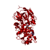
- Structure visualization
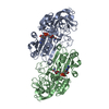
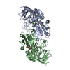
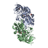
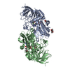
| Structure viewer | Molecule: MolmilJmol/JSmol |
|---|
- Downloads & links
-Download
| PDBx/mmCIF format | 1htb.cif.gz | 155.5 KB | Display | PDBx/mmCIF format |
|---|---|---|---|---|
| PDB format | pdb1htb.ent.gz | 122.3 KB | Display | PDB format |
| PDBx/mmJSON format | 1htb.json.gz | Tree view | PDBx/mmJSON format | |
| Others |  Other downloads Other downloads |
-Validation report
| Arichive directory | https://data.pdbj.org/pub/pdb/validation_reports/ht/1htbftp://data.pdbj.org/pub/pdb/validation_reports/ht/1htb | HTTPS FTP |
|---|
-Related structure data


-Links
 PDBj
PDBj

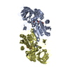
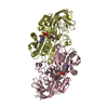
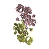
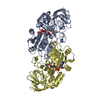
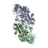
- Assembly
| Deposited unit | 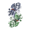
| ||||||||
|---|---|---|---|---|---|---|---|---|---|
| 1 |
| ||||||||
| Unit cell |
| ||||||||
| Atom site foot note | 1: CIS PROLINE - PRO A 62 / 2: CIS PROLINE - PRO B 62 | ||||||||
| Noncrystallographic symmetry (NCS) | NCS oper: (Code: given Matrix: (0.0737, 0.9799, -0.1853), Vector: |
-Components
-Protein , 1 types, 2 molecules AB
| #1: Protein | Mass: 39719.223 Da / Num. of mol.: 2 Source method: isolated from a genetically manipulated source Details: HOMODIMERIC WITH ONE NAD+ AND ONE 4-IODOPYRAZOLE PER SUBUNIT Source: (gene. exp.) Homo sapiens (human) / Gene: HUMAN BETA3 CDNA / Organ: LIVER / Plasmid: PKK223-3 / Gene (production host): HUMAN BETA3 CDNA / Production host:  |
|---|
-Non-polymers , 5 types, 151 molecules 


| #2: Chemical | ChemComp-ZN /  Mass: 65.409 Da / Num. of mol.: 4 / Source method: obtained synthetically / Formula: Zn Mass: 65.409 Da / Num. of mol.: 4 / Source method: obtained synthetically / Formula: Zn#3: Chemical |  Mass: 663.425 Da / Num. of mol.: 2 / Source method: obtained synthetically / Formula: C21H27N7O14P2 / Comment: NAD*YM Mass: 663.425 Da / Num. of mol.: 2 / Source method: obtained synthetically / Formula: C21H27N7O14P2 / Comment: NAD*YM#4: Chemical |  Mass: 193.974 Da / Num. of mol.: 2 / Source method: obtained synthetically / Formula: C3H3IN2 Mass: 193.974 Da / Num. of mol.: 2 / Source method: obtained synthetically / Formula: C3H3IN2#5: Chemical | ChemComp-CL / |  Mass: 35.453 Da / Num. of mol.: 1 / Source method: obtained synthetically / Formula: Cl Mass: 35.453 Da / Num. of mol.: 1 / Source method: obtained synthetically / Formula: Cl#6: Water | ChemComp-HOH / | Mass: 18.015 Da / Num. of mol.: 142 / Source method: isolated from a natural source / Formula: H2O |
|---|
-Experimental details
-Experiment
| Experiment | Method: X-RAY DIFFRACTION |
|---|
- Sample preparation
| Crystal | Density Matthews: 2.55 Å3/Da / Density % sol: 51.71 % | ||||||||||||||||||||||||||||||
|---|---|---|---|---|---|---|---|---|---|---|---|---|---|---|---|---|---|---|---|---|---|---|---|---|---|---|---|---|---|---|---|
| Crystal grow | *PLUS Temperature: 25 ℃ / pH: 7.5 / Method: vapor diffusion, sitting drop | ||||||||||||||||||||||||||||||
| Components of the solutions | *PLUS
|
-Data collection
| Diffraction source | Wavelength: 1.54 |
|---|---|
| Detector | Type: RIGAKU / Detector: IMAGE PLATE / Date: Feb 14, 1994 |
| Radiation | Monochromatic (M) / Laue (L): M / Scattering type: x-ray |
| Radiation wavelength | Wavelength: 1.54 Å / Relative weight: 1 |
| Reflection | Resolution: 2.37→47.97 Å / Num. obs: 26989 / % possible obs: 83.7 % / Redundancy: 2 % / Rmerge(I) obs: 0.1 |
| Reflection | *PLUS Highest resolution: 2.4 Å / Num. measured all: 53103 / Rmerge(I) obs: 0.1 |
| Reflection shell | *PLUS Highest resolution: 2.4 Å / Lowest resolution: 2.51 Å / % possible obs: 70 % |
- Processing
| Software |
| ||||||||||||||||||||||||||||||||||||||||||||||||||||||||||||
|---|---|---|---|---|---|---|---|---|---|---|---|---|---|---|---|---|---|---|---|---|---|---|---|---|---|---|---|---|---|---|---|---|---|---|---|---|---|---|---|---|---|---|---|---|---|---|---|---|---|---|---|---|---|---|---|---|---|---|---|---|---|
| Refinement | Resolution: 2.4→8 Å / σ(F): 1 Details: THE FOLLOWING DISTANCES ARE SIMILAR TO THOSE FOUND IN 1HDY (REFERENCE 1 ABOVE) AND ARE ALSO SIMILAR TO THE INTERACTION DISTANCES REPORTED BY EKLUND, H., SAMAMA, J.-P., WALLEN, L. (1982) ...Details: THE FOLLOWING DISTANCES ARE SIMILAR TO THOSE FOUND IN 1HDY (REFERENCE 1 ABOVE) AND ARE ALSO SIMILAR TO THE INTERACTION DISTANCES REPORTED BY EKLUND, H., SAMAMA, J.-P., WALLEN, L. (1982) PYRAZOLE BINDING IN CRYSTALLINE BINARY AND TERNARY COMPLEXES WITH LIVER ALCOHOL DEHYDROGENASE. BIOCHEMISTRY, VOL. 21, PP. 4858 - 4866) BETWEEN 4-IODOPYRAZOLE AND HORSE LIVER EE ADH (NO PDB ENTRY). SIMILAR CONTACT DISTANCES ARE ALSO FOUND IN 1DEH. 4-IODOPYRAZOLE FORMS A TRANSITION-STATE-LIKE COMPLEX WITH ADH AND EXHIBITS NEARLY COVALENT BOND CONTACT DISTANCES BETWEEN ITS TWO ADJACENT NITROGENS AND THE C4 ATOM OF NAD+ AND THE CATALYTIC ZINC ATOM: 377 A NAD NC4 378 A PYZ N2 HET-HET 1.7 376 B ZN ZN 378 B PYZ N1 HET-HET 2.0 377 B NAD NC4 378 B PYZ N2 HET-HET 2.1 376 A ZN ZN 378 A PYZ N1 HET-HET 2.3 RESIDUE ILE 368 IS AN OUTLIER IN THE RAMACHANDRAN PLOT. IT LIES IN THIS REGION OF THE RAMACHANDRAN PLOT IN ALL ADH STRUCTURES DEPOSITED IN THE DATA BANK. THUS, IT APPEARS THAT LOCAL STRUCTURAL FEATURES CONSTRAIN ITS CONFORMATION TO THIS POSITION. SER B 298 APPEARS TO OCCUPY AT LEAST TWO DISTINCT CONFORMATIONS. THE AUTHORS HAVE NOT ATTEMPTED TO MODEL POSITIONAL DISORDER, BUT ANY ONE CONFORMATION DOES NOT REFINE WELL TO THE OBSERVED DENSITY. THIS PROBLEM DOES NOT OCCUR IN THE A SUBUNIT.
| ||||||||||||||||||||||||||||||||||||||||||||||||||||||||||||
| Displacement parameters | Biso mean: 24.3 Å2 | ||||||||||||||||||||||||||||||||||||||||||||||||||||||||||||
| Refine analyze | Luzzati coordinate error obs: 0.23 Å | ||||||||||||||||||||||||||||||||||||||||||||||||||||||||||||
| Refinement step | Cycle: LAST / Resolution: 2.4→8 Å
| ||||||||||||||||||||||||||||||||||||||||||||||||||||||||||||
| Refine LS restraints |
| ||||||||||||||||||||||||||||||||||||||||||||||||||||||||||||
| Software | *PLUS Name: X-PLOR / Classification: refinement | ||||||||||||||||||||||||||||||||||||||||||||||||||||||||||||
| Refinement | *PLUS Highest resolution: 2.4 Å / Lowest resolution: 8 Å | ||||||||||||||||||||||||||||||||||||||||||||||||||||||||||||
| Solvent computation | *PLUS | ||||||||||||||||||||||||||||||||||||||||||||||||||||||||||||
| Displacement parameters | *PLUS | ||||||||||||||||||||||||||||||||||||||||||||||||||||||||||||
| Refine LS restraints | *PLUS
|
