 Movie
Movie Controller

[English] 日本語
 Yorodumi
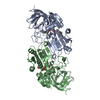
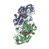
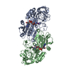
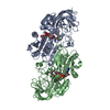
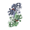
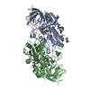
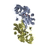
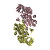
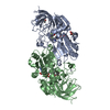
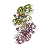
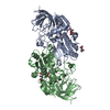
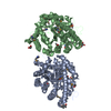
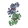
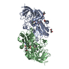
- PDB-1u3u: Crystal Structure of Human Alcohol Dehydrogenase Beta-1-Beta-1 Is... -
+ Open data

- Basic information
| Entry | Database: PDB / ID: 1u3u | ||||||
|---|---|---|---|---|---|---|---|
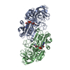
| Title | Crystal Structure of Human Alcohol Dehydrogenase Beta-1-Beta-1 Isoform Complexed with N-Benzylformamide Determined to 1.6 Angstrom Resolution | ||||||
 Components Components | Alcohol dehydrogenase beta chain | ||||||
 Keywords Keywords | OXIDOREDUCTASE | ||||||
| Function / homology |  Function and homology information Function and homology informationall-trans-retinol dehydrogenase (NAD+) / Ethanol oxidation / alcohol dehydrogenase (NAD+) activity / all-trans-retinol dehydrogenase (NAD+) activity / retinoic acid metabolic process / retinol metabolic process / ciliary transition zone / retinoid metabolic process / cilium / ciliary basal body ...all-trans-retinol dehydrogenase (NAD+) / Ethanol oxidation / alcohol dehydrogenase (NAD+) activity / all-trans-retinol dehydrogenase (NAD+) activity / retinoic acid metabolic process / retinol metabolic process / ciliary transition zone / retinoid metabolic process / cilium / ciliary basal body / zinc ion binding / nucleoplasm / plasma membrane / cytosol Similarity search - Function | ||||||
| Biological species |  Homo sapiens (human) Homo sapiens (human) | ||||||
| Method |  X-RAY DIFFRACTION / MOLECULAR REPLACEMENT / Resolution: 1.6 Å X-RAY DIFFRACTION / MOLECULAR REPLACEMENT / Resolution: 1.6 Å | ||||||
 Authors Authors | Gibbons, B.J. / Hurley, T.D. | ||||||
 Citation Citation | Journal: Biochemistry / Year: 2004 Title: Structure of three class I human alcohol dehydrogenases complexed with isoenzyme specific formamide inhibitors Authors: Gibbons, B.J. / Hurley, T.D. | ||||||
| History |
|
- Structure visualization
| Structure viewer | Molecule: MolmilJmol/JSmol |
|---|
- Downloads & links
-Download
| PDBx/mmCIF format | 1u3u.cif.gz | 182.6 KB | Display | PDBx/mmCIF format |
|---|---|---|---|---|
| PDB format | pdb1u3u.ent.gz | 139.2 KB | Display | PDB format |
| PDBx/mmJSON format | 1u3u.json.gz | Tree view | PDBx/mmJSON format | |
| Others |  Other downloads Other downloads |
-Validation report
| Arichive directory | https://data.pdbj.org/pub/pdb/validation_reports/u3/1u3uftp://data.pdbj.org/pub/pdb/validation_reports/u3/1u3u | HTTPS FTP |
|---|
-Related structure data
| Related structure data | 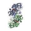 1u3tC 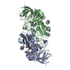 1u3vC  1u3wC 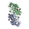 1hszS S: Starting model for refinement C: citing same article ( |
|---|---|
| Similar structure data |
-Links
 PDBj
PDBj

- Assembly
| Deposited unit | 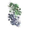
| ||||||||
|---|---|---|---|---|---|---|---|---|---|
| 1 |
| ||||||||
| Unit cell |
| ||||||||
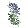
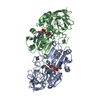
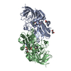
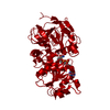
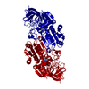
| Details | The assymetric unit contains one homodimer. |
-Components
-Protein , 1 types, 2 molecules AB
| #1: Protein | Mass: 39773.277 Da / Num. of mol.: 2 Source method: isolated from a genetically manipulated source Source: (gene. exp.) Homo sapiens (human) / Gene: ADH1B / Plasmid: pKK2233 / Production host:  |
|---|
-Non-polymers , 5 types, 1156 molecules 

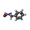


| #2: Chemical | ChemComp-ZN /  Mass: 65.409 Da / Num. of mol.: 4 / Source method: obtained synthetically / Formula: Zn Mass: 65.409 Da / Num. of mol.: 4 / Source method: obtained synthetically / Formula: Zn#3: Chemical |  Mass: 663.425 Da / Num. of mol.: 2 / Source method: obtained synthetically / Formula: C21H27N7O14P2 / Comment: NAD*YM Mass: 663.425 Da / Num. of mol.: 2 / Source method: obtained synthetically / Formula: C21H27N7O14P2 / Comment: NAD*YM#4: Chemical |  Mass: 135.163 Da / Num. of mol.: 2 / Source method: obtained synthetically / Formula: C8H9NO Mass: 135.163 Da / Num. of mol.: 2 / Source method: obtained synthetically / Formula: C8H9NO#5: Chemical | ChemComp-PO4 / |  Mass: 94.971 Da / Num. of mol.: 1 / Source method: obtained synthetically / Formula: PO4 Mass: 94.971 Da / Num. of mol.: 1 / Source method: obtained synthetically / Formula: PO4#6: Water | ChemComp-HOH / | Mass: 18.015 Da / Num. of mol.: 1147 / Source method: isolated from a natural source / Formula: H2O |
|---|
-Experimental details
-Experiment
| Experiment | Method: X-RAY DIFFRACTION / Number of used crystals: 1 |
|---|
- Sample preparation
| Crystal | Density Matthews: 2.4 Å3/Da / Density % sol: 49.1 % |
|---|---|
| Crystal grow | Temperature: 277 K / Method: vapor diffusion, sitting drop / pH: 7.5 Details: 15mg/ml enzyme, 2mM NAD+, 50mM sodium phosphate, and 11.5-13% PEG 8000, pH 7.5, VAPOR DIFFUSION, SITTING DROP, temperature 277K |
-Data collection
| Diffraction | Mean temperature: 100 K |
|---|---|
| Diffraction source | Source: ROTATING ANODE / Type: RIGAKU / Wavelength: 1.54059 Å |
| Detector | Type: RIGAKU RAXIS IIC / Detector: IMAGE PLATE / Date: Mar 1, 1999 |
| Radiation | Monochromator: YALE MIRRORS / Protocol: SINGLE WAVELENGTH / Monochromatic (M) / Laue (L): M / Scattering type: x-ray |
| Radiation wavelength | Wavelength: 1.54059 Å / Relative weight: 1 |
| Reflection | Resolution: 1.6→30 Å / Num. all: 88679 / Num. obs: 88679 / % possible obs: 90.7 % / Observed criterion σ(F): 0 / Observed criterion σ(I): 0 / Redundancy: 2.4 % / Biso Wilson estimate: 14.8 Å2 / Rmerge(I) obs: 0.046 / Net I/σ(I): 20.9 |
| Reflection shell | Resolution: 1.6→1.66 Å / Rmerge(I) obs: 0.172 / Mean I/σ(I) obs: 6.6 / Num. unique all: 8039 / % possible all: 82.4 |
- Processing
| Software |
| |||||||||||||||||||||||||
|---|---|---|---|---|---|---|---|---|---|---|---|---|---|---|---|---|---|---|---|---|---|---|---|---|---|---|
| Refinement | Method to determine structure: MOLECULAR REPLACEMENT Starting model: PDB ENTRY 1HSZ Resolution: 1.6→30 Å / Isotropic thermal model: overall / Cross valid method: THROUGHOUT / σ(F): 0 / σ(I): 0 / Stereochemistry target values: MAXIMUM LIKELIHOOD
| |||||||||||||||||||||||||
| Displacement parameters | Biso mean: 14.8 Å2 | |||||||||||||||||||||||||
| Refine analyze |
| |||||||||||||||||||||||||
| Refinement step | Cycle: LAST / Resolution: 1.6→30 Å
| |||||||||||||||||||||||||
| Refine LS restraints |
|
