 Movie
Movie Controller

[English] 日本語
 Yorodumi
- PDB-1axe: CRYSTAL STRUCTURE OF THE ACTIVE-SITE MUTANT PHE93->TRP OF HORSE L... -
+ Open data

- Basic information
| Entry | Database: PDB / ID: 1axe | ||||||
|---|---|---|---|---|---|---|---|
| Title | CRYSTAL STRUCTURE OF THE ACTIVE-SITE MUTANT PHE93->TRP OF HORSE LIVER ALCOHOL DEHYDROGENASE IN COMPLEX WITH NAD AND INHIBITOR TRIFLUOROETHANOL | ||||||
 Components Components | ALCOHOL DEHYDROGENASE | ||||||
 Keywords Keywords | OXIDOREDUCTASE / OXIDOREDUCTASE (NAD(A)-CHOH(D)) / ALCOHOL DEHYDROGENASE | ||||||
| Function / homology |  Function and homology information Function and homology informationall-trans-retinol dehydrogenase (NAD+) activity / alcohol dehydrogenase / retinoic acid metabolic process / retinol metabolic process / zinc ion binding / cytosol Similarity search - Function | ||||||
| Biological species |  | ||||||
| Method |  X-RAY DIFFRACTION / MOLECULAR REPLACEMENT / Resolution: 2 Å X-RAY DIFFRACTION / MOLECULAR REPLACEMENT / Resolution: 2 Å | ||||||
 Authors Authors | Colby, T.D. / Chin, J.K. / Goldstein, B.M. | ||||||
 Citation Citation | Journal: Proc.Natl.Acad.Sci.USA / Year: 1997 Title: A link between protein structure and enzyme catalyzed hydrogen tunneling. Authors: Bahnson, B.J. / Colby, T.D. / Chin, J.K. / Goldstein, B.M. / Klinman, J.P. #1: Journal: Acta Crystallogr.,Sect.D / Year: 1994Title: Refined Crystal Structure of Liver Alcohol Dehydrogenase-Nadh Complex at 1.8 A Resolution Authors: Al-Karadaghi, S. / Cedergren-Zeppezauer, E.S. / Hovmoller, S. / Petratos, K. / Terry, H. / Wilson, K.S. #2: Journal: J.Biol.Chem. / Year: 1986Title: Interdomain Motion in Liver Alcohol Dehydrogenase. Structural and Energetic Analysis of the Hinge Bending Mode Authors: Colonna-Cesari, F. / Perahia, D. / Karplus, M. / Eklund, H. / Branden, C.I. / Tapia, O. #3: Journal: Biochemistry / Year: 1984Title: Crystallographic Investigations of Nicotinamide Adenine Dinucleotide Binding to Horse Liver Alcohol Dehydrogenase Authors: Eklund, H. / Samama, J.P. / Jones, T.A. #4: Journal: Biochemistry / Year: 1983Title: Crystal-Structure Determination of Reduced Nicotinamide Adenine Dinucleotide Complex with Horse Liver Alcohol Dehydrogenase Maintained in its Apo Conformation by Zinc-Bound Imidazole Authors: Cedergren-Zeppezauer, E. #5: Journal: J.Biol.Chem. / Year: 1983Title: Three-Dimensional Structure of Isonicotinimidylated Liver Alcohol Dehydrogenase Authors: Plapp, B.V. / Eklund, H. / Jones, T.A. / Branden, C.I. #6: Journal: Proc.Natl.Acad.Sci.USA / Year: 1983Title: Crystal Structures of the Active Site in Specifically Metal-Depleted and Cobalt-Substituted Horse Liver Alcohol Dehydrogenase Derivatives Authors: Schneider, G. / Eklund, H. / Cedergren-Zeppezauer, E. / Zeppezauer, M. #7: Journal: Biochemistry / Year: 1982Title: Pyrazole Binding in Crystalline Binary and Ternary Complexes with Liver Alcohol Dehydrogenase Authors: Eklund, H. / Samama, J.P. / Wallen, L. #8: Journal: Biochemistry / Year: 1982Title: Crystal Structure Determinations of Coenzyme Analogue and Substrate Complexes of Liver Alcohol Dehydrogenase. Binding of 1,4,5,6-Tetrahydronicotinamide Adenine Dinucleotide and Trans-4-(N,N- ...Title: Crystal Structure Determinations of Coenzyme Analogue and Substrate Complexes of Liver Alcohol Dehydrogenase. Binding of 1,4,5,6-Tetrahydronicotinamide Adenine Dinucleotide and Trans-4-(N,N-Dimethylamino)Cinnamaldehyde to the Enzyme Authors: Cedergren-Zeppezauer, E. / Samama, J.P. / Eklund, H. #9: Journal: J.Biol.Chem. / Year: 1982Title: Binding of Substrate in a Ternary Complex of Horse Liver Alcohol Dehydrogenase Authors: Eklund, H. / Plapp, B.V. / Samama, J.P. / Branden, C.I. #10: Journal: J.Biol.Chem. / Year: 1979Title: Structural Differences between Apo-and Holoenzyme of Horse Liver Alcohol Dehydrogenase Authors: Eklund, H. / Branden, C.I. #11: Journal: J.Mol.Biol. / Year: 1976Title: Three-Dimensional Structure of Horse Liver Alcohol Dehydrogenase at 2.4 A Resolution Authors: Eklund, H. / Nordstrom, B. / Zeppezauer, E. / Soderlund, G. / Ohlsson, I. / Boiwe, T. / Soderberg, B.O. / Tapia, O. / Branden, C.I. / Akeson, A. | ||||||
| History |
|
- Structure visualization
| Structure viewer | Molecule: MolmilJmol/JSmol |
|---|
- Downloads & links
-Download
| PDBx/mmCIF format | 1axe.cif.gz | 155.1 KB | Display | PDBx/mmCIF format |
|---|---|---|---|---|
| PDB format | pdb1axe.ent.gz | 122 KB | Display | PDB format |
| PDBx/mmJSON format | 1axe.json.gz | Tree view | PDBx/mmJSON format | |
| Others |  Other downloads Other downloads |
-Validation report
| Arichive directory | https://data.pdbj.org/pub/pdb/validation_reports/ax/1axeftp://data.pdbj.org/pub/pdb/validation_reports/ax/1axe | HTTPS FTP |
|---|
-Related structure data
| Related structure data |  1axgC  2ohxS S: Starting model for refinement C: citing same article ( |
|---|---|
| Similar structure data |

-Links
 PDBj
PDBj

- Assembly
| Deposited unit | 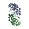
| ||||||||
|---|---|---|---|---|---|---|---|---|---|
| 1 |
| ||||||||
| Unit cell |
| ||||||||
| Noncrystallographic symmetry (NCS) | NCS oper: (Code: given Matrix: (0.122993, 0.984919, 0.121686), Vector: |
-Components
| #1: Protein | Mass: 39892.309 Da / Num. of mol.: 2 / Mutation: F93W Source method: isolated from a genetically manipulated source Details: HOLO-ENZYME / Source: (gene. exp.)  #2: Chemical | ChemComp-ZN /   Mass: 65.409 Da / Num. of mol.: 4 / Source method: obtained synthetically / Formula: Zn Mass: 65.409 Da / Num. of mol.: 4 / Source method: obtained synthetically / Formula: Zn#3: Chemical |   Mass: 663.425 Da / Num. of mol.: 2 / Source method: obtained synthetically / Formula: C21H27N7O14P2 / Comment: NAD*YM Mass: 663.425 Da / Num. of mol.: 2 / Source method: obtained synthetically / Formula: C21H27N7O14P2 / Comment: NAD*YM#4: Chemical |   Mass: 100.040 Da / Num. of mol.: 2 / Source method: obtained synthetically / Formula: C2H3F3O Mass: 100.040 Da / Num. of mol.: 2 / Source method: obtained synthetically / Formula: C2H3F3O#5: Water | ChemComp-HOH / |  Mass: 18.015 Da / Num. of mol.: 116 / Source method: isolated from a natural source / Formula: H2O Mass: 18.015 Da / Num. of mol.: 116 / Source method: isolated from a natural source / Formula: H2O |
|---|
-Experimental details
-Experiment
| Experiment | Method: X-RAY DIFFRACTION / Number of used crystals: 4 |
|---|
- Sample preparation
| Crystal | Density Matthews: 2.5 Å3/Da / Density % sol: 49.98 % | ||||||||||||||||||||||||||||||||||||||||||||||||
|---|---|---|---|---|---|---|---|---|---|---|---|---|---|---|---|---|---|---|---|---|---|---|---|---|---|---|---|---|---|---|---|---|---|---|---|---|---|---|---|---|---|---|---|---|---|---|---|---|---|
| Crystal grow | Temperature: 277 K / Method: vapor diffusion, hanging drop / pH: 8.4 Details: CRYSTALS WERE GROWN FROM HANGING DROPS. THE INITIAL DROP CONTAINING 16MG/ML PROTEIN, A 10X EXCESS OF NAD, 5MM TRIFLUOROETHANOL AND 5% V/V OF PEG400 IN 50MM TRIS-HCL PH 8.4 WERE EQUILIBRATED ...Details: CRYSTALS WERE GROWN FROM HANGING DROPS. THE INITIAL DROP CONTAINING 16MG/ML PROTEIN, A 10X EXCESS OF NAD, 5MM TRIFLUOROETHANOL AND 5% V/V OF PEG400 IN 50MM TRIS-HCL PH 8.4 WERE EQUILIBRATED AT 4 DEGREES C OVER WELLS OF SIMILAR COMPOSITION CONTAINING 15-17% PEG400., vapor diffusion - hanging drop, temperature 277K | ||||||||||||||||||||||||||||||||||||||||||||||||
| Crystal | *PLUS Density % sol: 51 % | ||||||||||||||||||||||||||||||||||||||||||||||||
| Crystal grow | *PLUS Temperature: 4 ℃ / Method: vapor diffusion | ||||||||||||||||||||||||||||||||||||||||||||||||
| Components of the solutions | *PLUS
|
-Data collection
| Diffraction | Mean temperature: 277 K |
|---|---|
| Diffraction source | Source: ROTATING ANODE / Type: RIGAKU RUH2R / Wavelength: 1.5418 |
| Detector | Type: RIGAKU RAXIS II / Detector: IMAGE PLATE / Date: Sep 17, 1995 / Details: MONOCHROMATOR |
| Radiation | Monochromator: NI FILTER / Monochromatic (M) / Laue (L): M / Scattering type: x-ray |
| Radiation wavelength | Wavelength: 1.5418 Å / Relative weight: 1 |
| Reflection | Resolution: 2→99 Å / Num. obs: 43756 / % possible obs: 85.2 % / Observed criterion σ(I): 2 / Redundancy: 1.83 % / Rmerge(I) obs: 0.064 / Rsym value: 0.064 / Net I/σ(I): 5 |
| Reflection shell | Resolution: 2→2.07 Å / Redundancy: 1.6 % / Rmerge(I) obs: 0.31 / Mean I/σ(I) obs: 1.3 / Rsym value: 0.31 / % possible all: 64.6 |
- Processing
| Software |
| ||||||||||||||||||||||||||||||||||||||||||||||||||||||||||||
|---|---|---|---|---|---|---|---|---|---|---|---|---|---|---|---|---|---|---|---|---|---|---|---|---|---|---|---|---|---|---|---|---|---|---|---|---|---|---|---|---|---|---|---|---|---|---|---|---|---|---|---|---|---|---|---|---|---|---|---|---|---|
| Refinement | Method to determine structure: MOLECULAR REPLACEMENT Starting model: PDB ENTRY 2OHX Resolution: 2→10 Å / Rfactor Rfree error: 0.006 / Data cutoff high absF: 100000 / Data cutoff low absF: 0.1 / Isotropic thermal model: RESTRAINED / Cross valid method: THROUGHOUT / σ(F): 2
| ||||||||||||||||||||||||||||||||||||||||||||||||||||||||||||
| Refinement step | Cycle: LAST / Resolution: 2→10 Å
| ||||||||||||||||||||||||||||||||||||||||||||||||||||||||||||
| Refine LS restraints |
| ||||||||||||||||||||||||||||||||||||||||||||||||||||||||||||
| Xplor file |
| ||||||||||||||||||||||||||||||||||||||||||||||||||||||||||||
| Software | *PLUS Name: X-PLOR / Version: 3.1 / Classification: refinement | ||||||||||||||||||||||||||||||||||||||||||||||||||||||||||||
| Refine LS restraints | *PLUS
|
