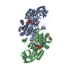
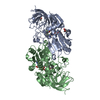
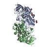
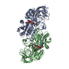
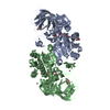
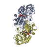
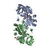
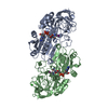
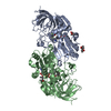
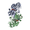
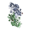
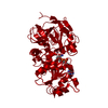
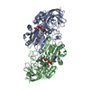
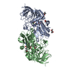
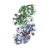
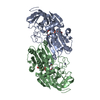
Entry Database : PDB / ID : 4xd2Title Horse liver alcohol dehydrogenase-NADH complex Alcohol dehydrogenase E chain Keywords / / / Function / homology Function Domain/homology Component
/ / / / / / / / / / / / / / / / / / / / / / / / / / / / / / Biological species Equus caballus (horse)Method / / / Resolution : 1.1 Å Authors Plapp, B.V. / Baskar Raj, S. / Ferraro, D.J. Funding support Organization Grant number Country National Institutes of Health/National Institute on Alcohol Abuse and Alcoholism (NIH/NIAAA) AA00279 National Institutes of Health/National Institute of General Medical Sciences (NIH/NIGMS) T32 GM008365
Journal : To Be Published Title : Structure of Horse liver alcohol dehydrogenase complexed with NADHAuthors : Plapp, B.V. / Baskar Raj, S. / Ferraro, D.J. History Deposition Dec 18, 2014 Deposition site / Processing site Revision 1.0 Jan 14, 2015 Provider / Type Revision 2.0 Nov 22, 2017 Group Advisory / Atomic model ... Advisory / Atomic model / Author supporting evidence / Derived calculations / Source and taxonomy Category atom_site / atom_site_anisotrop ... atom_site / atom_site_anisotrop / entity_src_nat / pdbx_audit_support / pdbx_distant_solvent_atoms / pdbx_struct_conn_angle / pdbx_struct_oper_list / struct_conn / struct_site_gen Item _atom_site.B_iso_or_equiv / _atom_site.Cartn_x ... _atom_site.B_iso_or_equiv / _atom_site.Cartn_x / _atom_site.Cartn_y / _atom_site.Cartn_z / _atom_site.auth_seq_id / _atom_site.label_alt_id / _atom_site.occupancy / _atom_site_anisotrop.U[1][1] / _atom_site_anisotrop.U[1][2] / _atom_site_anisotrop.U[1][3] / _atom_site_anisotrop.U[2][2] / _atom_site_anisotrop.U[2][3] / _atom_site_anisotrop.U[3][3] / _atom_site_anisotrop.pdbx_auth_seq_id / _atom_site_anisotrop.pdbx_label_alt_id / _entity_src_nat.pdbx_alt_source_flag / _pdbx_audit_support.funding_organization / _pdbx_distant_solvent_atoms.auth_seq_id / _pdbx_struct_conn_angle.ptnr3_auth_seq_id / _pdbx_struct_oper_list.symmetry_operation / _struct_conn.ptnr2_auth_seq_id / _struct_site_gen.auth_seq_id Revision 2.1 Dec 11, 2019 Group / Category / Item Revision 2.2 Sep 27, 2023 Group Data collection / Database references ... Data collection / Database references / Derived calculations / Refinement description Category chem_comp_atom / chem_comp_bond ... chem_comp_atom / chem_comp_bond / database_2 / pdbx_initial_refinement_model / struct_conn Item _database_2.pdbx_DOI / _database_2.pdbx_database_accession ... _database_2.pdbx_DOI / _database_2.pdbx_database_accession / _struct_conn.pdbx_dist_value / _struct_conn.pdbx_ptnr1_label_alt_id / _struct_conn.pdbx_ptnr2_label_alt_id / _struct_conn.ptnr1_auth_asym_id / _struct_conn.ptnr1_auth_comp_id / _struct_conn.ptnr1_auth_seq_id / _struct_conn.ptnr1_label_asym_id / _struct_conn.ptnr1_label_atom_id / _struct_conn.ptnr1_label_comp_id / _struct_conn.ptnr1_label_seq_id / _struct_conn.ptnr2_auth_asym_id / _struct_conn.ptnr2_auth_comp_id / _struct_conn.ptnr2_auth_seq_id / _struct_conn.ptnr2_label_asym_id / _struct_conn.ptnr2_label_atom_id / _struct_conn.ptnr2_label_comp_id
Show all Show less
 Movie
Movie Controller


 Open data
 Basic information
 Components
 Keywords
 Function and homology information
 X-RAY DIFFRACTION /
  Authors
 United States, 2items
  Citation
 Structure visualization
 Downloads & links
 Other downloads
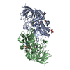


 PDBj
PDBj


 Assembly
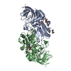

 Mass: 65.409 Da / Num. of mol.: 4 / Source method: obtained synthetically / Formula: Zn
 Mass: 665.441 Da / Num. of mol.: 2 / Source method: obtained synthetically / Formula: C21H29N7O14P2
 Mass: 118.174 Da / Num. of mol.: 3 / Source method: obtained synthetically / Formula: C6H14O2 / Comment: precipitant*YM
 Mass: 18.015 Da / Num. of mol.: 804 / Source method: isolated from a natural source / Formula: H2O
 Sample preparation
 Processing