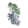
 Movie
Movie Controller

[English] 日本語
 Yorodumi
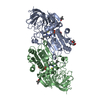
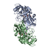
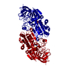
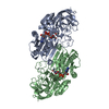
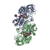
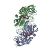
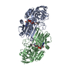
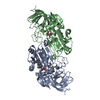
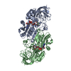
- PDB-1n8k: Horse Liver Alcohol Dehydrogenase Val292Thr Mutant Complexed to N... -
+ Open data

- Basic information
| Entry | Database: PDB / ID: 1n8k | ||||||
|---|---|---|---|---|---|---|---|
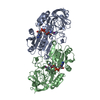
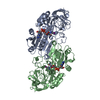
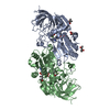
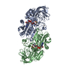
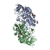
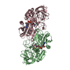
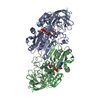
| Title | Horse Liver Alcohol Dehydrogenase Val292Thr Mutant Complexed to NAD+ and Pyrazole | ||||||
 Components Components | Alcohol Dehydrogenase E chain | ||||||
 Keywords Keywords | OXIDOREDUCTASE / DEHYDROGENASE / ALCOHOL / NICOTINAMIDE COENZYME / MUTANT / PYRAZOLE | ||||||
| Function / homology |  Function and homology information Function and homology informationall-trans-retinol dehydrogenase (NAD+) activity / alcohol dehydrogenase / retinoic acid metabolic process / retinol metabolic process / zinc ion binding / cytosol Similarity search - Function | ||||||
| Biological species |  | ||||||
| Method |  X-RAY DIFFRACTION / SYNCHROTRON / MOLECULAR REPLACEMENT / Resolution: 1.13 Å X-RAY DIFFRACTION / SYNCHROTRON / MOLECULAR REPLACEMENT / Resolution: 1.13 Å | ||||||
 Authors Authors | Rubach, J.K. / Plapp, B.V. | ||||||
 Citation Citation | Journal: Biochemistry / Year: 2003 Title: Amino Acid Residues in the Nicotinamide Binding Site Contribute to Catalysis by Horse Liver Alcohol Dehydrogenase Authors: Rubach, J.K. / Plapp, B.V. #1: Journal: Biochemistry / Year: 1982Title: Pyrazole Binding in Crystalline Binary and Ternary Complexes with Liver Alcohol Dehydrogenase Authors: Eklund, H. / Samama, J.P. / Wallen, L. #2: Journal: J.Biol.Chem. / Year: 1996Title: X-Ray Structure of Human beta3beta3 Alcohol Dehydrogenase. The contribution of ionic interactions to coenzyme binding. Authors: Davis, G.J. / Bosron, W.F. / Stone, C.L. / Owusu-Dekyi, K. / Hurley, T.D. #3: Journal: J.Mol.Biol. / Year: 1994Title: Structures of Three Human Beta Alcohol Dehydrogenase Variants. Correlations with their Functional Differences. Authors: Hurley, T.D. / Bosron, W.F. / Stone, C.L. / Amzel, L.M. #4: Journal: J.Mol.Biol. / Year: 1981Title: Structure of A Triclinic Ternary Complex of Horse Liver Alcohol Dehydrogenase at 2.9 A Resolution Authors: Eklund, H. / Samama, J.P. / Wallen, L. / Branden, C.I. / Akeson, A. / Jones, T.A. #5: Journal: J.Mol.Biol. / Year: 1976Title: Three-Dimensional Structure of Horse Liver Alcohol Dehydrogenase at 2.4 A Resolution Authors: Eklund, H. / Nordstrom, B. / Zeppezauer, E. / Soderlund, G. / Ohlsson, I. / Boiwe, T. / Soderberg, B.O. / Tapia, O. / Branden, C.I. / Akeson, A. | ||||||
| History |
| ||||||
| Remark 600 | HETEROGEN THE STRUCTURE DETERMINED IN THIS ENTRY CONTAINS THE HETEROATOM MOLECULE NAJ, WHICH IS ...HETEROGEN THE STRUCTURE DETERMINED IN THIS ENTRY CONTAINS THE HETEROATOM MOLECULE NAJ, WHICH IS OXIDIZED NICOTINAMIDE ADENINE DINUCLEOTIDE, BUT THE RESTRAINTS ON THE PLANARITY OF THE NICOTINAMIDE RING WERE REMOVED DURING REFINEMENT WITH THE RESULT THAT THE RING IS PUCKERED. | ||||||
| Remark 285 | CRYST1 TO GENERATE THE STANDARD UNIT CELL FOR P1 FROM THE CELL DESCRIBED IN THE CRYST CARD, APPLY ...CRYST1 TO GENERATE THE STANDARD UNIT CELL FOR P1 FROM THE CELL DESCRIBED IN THE CRYST CARD, APPLY THE FOLLOWING: 0.000000 1.000000 0.000000 0.00000 0.000000 0.000000 1.000000 0.00000 1.000000 0.000000 0.000000 0.00000 WHICH YIELDS THE FOLLOWING UNIT CELL PARAMETERS: 51.4 92.7 44.3 103.0 109.9 91.9 P 1 |
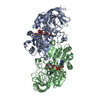
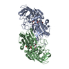
- Structure visualization
| Structure viewer | Molecule: MolmilJmol/JSmol |
|---|
- Downloads & links
-Download
| PDBx/mmCIF format | 1n8k.cif.gz | 305.6 KB | Display | PDBx/mmCIF format |
|---|---|---|---|---|
| PDB format | pdb1n8k.ent.gz | 245.6 KB | Display | PDB format |
| PDBx/mmJSON format | 1n8k.json.gz | Tree view | PDBx/mmJSON format | |
| Others |  Other downloads Other downloads |
-Validation report
| Arichive directory | https://data.pdbj.org/pub/pdb/validation_reports/n8/1n8kftp://data.pdbj.org/pub/pdb/validation_reports/n8/1n8k | HTTPS FTP |
|---|
-Related structure data
| Related structure data | 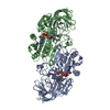 1n92C 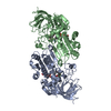 1hldS S: Starting model for refinement C: citing same article ( |
|---|---|
| Similar structure data |
-Links
 PDBj
PDBj

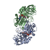
- Assembly
| Deposited unit | 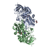
| ||||||||
|---|---|---|---|---|---|---|---|---|---|
| 1 |
| ||||||||
| Unit cell |
|
-Components
-Protein , 1 types, 2 molecules AB
| #1: Protein | Mass: 39855.246 Da / Num. of mol.: 2 / Mutation: V292T Source method: isolated from a genetically manipulated source Source: (gene. exp.)  |
|---|
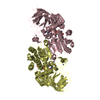
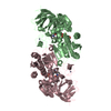
-Non-polymers , 5 types, 716 molecules 
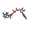


| #2: Chemical | ChemComp-ZN /  Mass: 65.409 Da / Num. of mol.: 4 / Source method: obtained synthetically / Formula: Zn Mass: 65.409 Da / Num. of mol.: 4 / Source method: obtained synthetically / Formula: Zn#3: Chemical |  Mass: 663.425 Da / Num. of mol.: 2 / Source method: obtained synthetically / Formula: C21H27N7O14P2 Mass: 663.425 Da / Num. of mol.: 2 / Source method: obtained synthetically / Formula: C21H27N7O14P2#4: Chemical |  Mass: 68.077 Da / Num. of mol.: 2 / Source method: obtained synthetically / Formula: C3H4N2 Mass: 68.077 Da / Num. of mol.: 2 / Source method: obtained synthetically / Formula: C3H4N2#5: Chemical | ChemComp-MPD / ( |  Mass: 118.174 Da / Num. of mol.: 1 / Source method: obtained synthetically / Formula: C6H14O2 / Comment: precipitant*YM Mass: 118.174 Da / Num. of mol.: 1 / Source method: obtained synthetically / Formula: C6H14O2 / Comment: precipitant*YM#6: Water | ChemComp-HOH / | Mass: 18.015 Da / Num. of mol.: 707 / Source method: isolated from a natural source / Formula: H2O |
|---|
-Experimental details
-Experiment
| Experiment | Method: X-RAY DIFFRACTION / Number of used crystals: 1 |
|---|
- Sample preparation
| Crystal | Density Matthews: 2.13 Å3/Da / Density % sol: 41.89 % | ||||||||||||||||||||||||||||||||||||||||||
|---|---|---|---|---|---|---|---|---|---|---|---|---|---|---|---|---|---|---|---|---|---|---|---|---|---|---|---|---|---|---|---|---|---|---|---|---|---|---|---|---|---|---|---|
| Crystal grow | Temperature: 277 K / pH: 7 / Details: MPD, pH 7.0, dialysis, temperature 277K, pH 7.00 | ||||||||||||||||||||||||||||||||||||||||||
| Crystal grow | *PLUS Temperature: 25 ℃ / Method: microdialysis | ||||||||||||||||||||||||||||||||||||||||||
| Components of the solutions | *PLUS
|
-Data collection
| Diffraction | Mean temperature: 100 K |
|---|---|
| Diffraction source | Source: SYNCHROTRON / Site: APS  / Beamline: 17-ID / Wavelength: 1 / Beamline: 17-ID / Wavelength: 1 |
| Detector | Type: MARRESEARCH / Detector: CCD / Date: Dec 9, 2001 |
| Radiation | Protocol: SINGLE WAVELENGTH / Monochromatic (M) / Laue (L): M / Scattering type: x-ray |
| Radiation wavelength | Wavelength: 1 Å / Relative weight: 1 |
| Reflection | Resolution: 1.13→20 Å / Num. obs: 218390 / Observed criterion σ(I): 0 / Redundancy: 2.2 % / Rsym value: 0.058 / Net I/σ(I): 32 |
| Reflection shell | Resolution: 1.13→1.16 Å / Rmerge(I) obs: 0.172 / Mean I/σ(I) obs: 6.4 / Rsym value: 0.172 / % possible all: 46.2 |
| Reflection | *PLUS Highest resolution: 1.13 Å / Lowest resolution: 20 Å / % possible obs: 81.6 % / Num. measured all: 6143593 / Rmerge(I) obs: 0.058 |
| Reflection shell | *PLUS % possible obs: 46.2 % / Rmerge(I) obs: 0.167 / Mean I/σ(I) obs: 6.4 |
- Processing
| Software |
| ||||||||||||||||||||||||||||||||||||||||||||||||||||||||||||||||||||||||||||||||||||||||||
|---|---|---|---|---|---|---|---|---|---|---|---|---|---|---|---|---|---|---|---|---|---|---|---|---|---|---|---|---|---|---|---|---|---|---|---|---|---|---|---|---|---|---|---|---|---|---|---|---|---|---|---|---|---|---|---|---|---|---|---|---|---|---|---|---|---|---|---|---|---|---|---|---|---|---|---|---|---|---|---|---|---|---|---|---|---|---|---|---|---|---|---|
| Refinement | Method to determine structure: MOLECULAR REPLACEMENT Starting model: PDB ENTRY 1HLD Resolution: 1.13→20 Å / Cor.coef. Fo:Fc: 0.98 / Cor.coef. Fo:Fc free: 0.975 / SU B: 0.489 / SU ML: 0.023 / Cross valid method: THROUGHOUT / σ(I): 0 / ESU R: 0.036 / ESU R Free: 0.036 / Stereochemistry target values: MAXIMUM LIKELIHOOD / Details: HYDROGENS HAVE BEEN ADDED IN THE RIDING POSITIONS
| ||||||||||||||||||||||||||||||||||||||||||||||||||||||||||||||||||||||||||||||||||||||||||
| Solvent computation | Ion probe radii: 0.8 Å / Shrinkage radii: 0.8 Å / VDW probe radii: 1.4 Å / Solvent model: BABINET MODEL WITH MASK | ||||||||||||||||||||||||||||||||||||||||||||||||||||||||||||||||||||||||||||||||||||||||||
| Displacement parameters | Biso mean: 18.194 Å2
| ||||||||||||||||||||||||||||||||||||||||||||||||||||||||||||||||||||||||||||||||||||||||||
| Refine analyze |
| ||||||||||||||||||||||||||||||||||||||||||||||||||||||||||||||||||||||||||||||||||||||||||
| Refinement step | Cycle: LAST / Resolution: 1.13→20 Å
| ||||||||||||||||||||||||||||||||||||||||||||||||||||||||||||||||||||||||||||||||||||||||||
| Refine LS restraints |
| ||||||||||||||||||||||||||||||||||||||||||||||||||||||||||||||||||||||||||||||||||||||||||
| LS refinement shell | Resolution: 1.131→1.161 Å / Total num. of bins used: 20 /
| ||||||||||||||||||||||||||||||||||||||||||||||||||||||||||||||||||||||||||||||||||||||||||
| Refinement | *PLUS Lowest resolution: 20 Å / Rfactor Rfree: 0.167 / Rfactor Rwork: 0.145 | ||||||||||||||||||||||||||||||||||||||||||||||||||||||||||||||||||||||||||||||||||||||||||
| Solvent computation | *PLUS | ||||||||||||||||||||||||||||||||||||||||||||||||||||||||||||||||||||||||||||||||||||||||||
| Displacement parameters | *PLUS | ||||||||||||||||||||||||||||||||||||||||||||||||||||||||||||||||||||||||||||||||||||||||||
| Refine LS restraints | *PLUS
| ||||||||||||||||||||||||||||||||||||||||||||||||||||||||||||||||||||||||||||||||||||||||||
| LS refinement shell | *PLUS Rfactor Rfree: 0.19 |
