 Movie
Movie Controller


+ Open data

- Basic information
| Entry | Database: PDB / ID: 1hso | ||||||
|---|---|---|---|---|---|---|---|
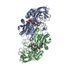
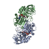
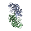
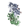
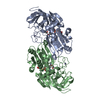
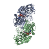
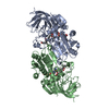
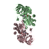
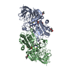
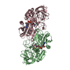
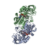
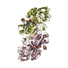
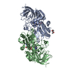
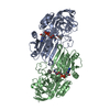
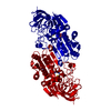
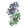
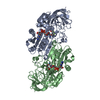
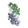
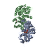
| Title | HUMAN ALPHA ALCOHOL DEHYDROGENASE (ADH1A) | ||||||
 Components Components | CLASS I ALCOHOL DEHYDROGENASE 1, ALPHA SUBUNIT | ||||||
 Keywords Keywords | OXIDOREDUCTASE / rossmann fold / alcohol dehydrogenase | ||||||
| Function / homology |  Function and homology information Function and homology informationbutanol dehydrogenase (NAD+) activity / Abacavir metabolism / alcohol metabolic process / Ethanol oxidation / RA biosynthesis pathway / alcohol dehydrogenase (NAD+) activity / all-trans-retinol dehydrogenase (NAD+) activity / alcohol dehydrogenase / retinoic acid metabolic process / retinol metabolic process ...butanol dehydrogenase (NAD+) activity / Abacavir metabolism / alcohol metabolic process / Ethanol oxidation / RA biosynthesis pathway / alcohol dehydrogenase (NAD+) activity / all-trans-retinol dehydrogenase (NAD+) activity / alcohol dehydrogenase / retinoic acid metabolic process / retinol metabolic process / zinc ion binding / cytosol Similarity search - Function | ||||||
| Biological species |  Homo sapiens (human) Homo sapiens (human) | ||||||
| Method |  X-RAY DIFFRACTION / MOLECULAR REPLACEMENT / Resolution: 2.5 Å X-RAY DIFFRACTION / MOLECULAR REPLACEMENT / Resolution: 2.5 Å | ||||||
 Authors Authors | Niederhut, M.S. / Gibbons, B.J. / Perez-Miller, S. / Hurley, T.D. | ||||||
 Citation Citation | Journal: Protein Sci. / Year: 2001 Title: Three-dimensional structures of the three human class I alcohol dehydrogenases. Authors: Niederhut, M.S. / Gibbons, B.J. / Perez-Miller, S. / Hurley, T.D. | ||||||
| History |
|
- Structure visualization
| Structure viewer | Molecule: MolmilJmol/JSmol |
|---|
- Downloads & links
-Download
| PDBx/mmCIF format | 1hso.cif.gz | 161.5 KB | Display | PDBx/mmCIF format |
|---|---|---|---|---|
| PDB format | pdb1hso.ent.gz | 125.5 KB | Display | PDB format |
| PDBx/mmJSON format | 1hso.json.gz | Tree view | PDBx/mmJSON format | |
| Others |  Other downloads Other downloads |
-Validation report
| Arichive directory | https://data.pdbj.org/pub/pdb/validation_reports/hs/1hsoftp://data.pdbj.org/pub/pdb/validation_reports/hs/1hso | HTTPS FTP |
|---|
-Related structure data
| Related structure data | 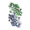 1hszC  1ht0C 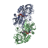 1dehS S: Starting model for refinement C: citing same article ( |
|---|---|
| Similar structure data |

-Links
 PDBj
PDBj


- Assembly
| Deposited unit | 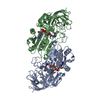
| ||||||||||
|---|---|---|---|---|---|---|---|---|---|---|---|
| 1 |
| ||||||||||
| Unit cell |
| ||||||||||
| Details | the biological assembly is the dimer present in the asymmetric unit |
-Components
| #1: Protein | Mass: 39776.469 Da / Num. of mol.: 2 / Fragment: ALPHA SUBUNIT Source method: isolated from a genetically manipulated source Source: (gene. exp.) Homo sapiens (human) / Gene: ADH1A / Plasmid: PKK223-3 / Production host:  #2: Chemical | ChemComp-ZN /   Mass: 65.409 Da / Num. of mol.: 4 / Source method: obtained synthetically / Formula: Zn Mass: 65.409 Da / Num. of mol.: 4 / Source method: obtained synthetically / Formula: Zn#3: Chemical |   Mass: 663.425 Da / Num. of mol.: 2 / Source method: obtained synthetically / Formula: C21H27N7O14P2 / Comment: NAD*YM Mass: 663.425 Da / Num. of mol.: 2 / Source method: obtained synthetically / Formula: C21H27N7O14P2 / Comment: NAD*YM#4: Chemical |   Mass: 193.974 Da / Num. of mol.: 2 / Source method: obtained synthetically / Formula: C3H3IN2 Mass: 193.974 Da / Num. of mol.: 2 / Source method: obtained synthetically / Formula: C3H3IN2#5: Water | ChemComp-HOH / |  Mass: 18.015 Da / Num. of mol.: 362 / Source method: isolated from a natural source / Formula: H2O Mass: 18.015 Da / Num. of mol.: 362 / Source method: isolated from a natural source / Formula: H2O |
|---|
-Experimental details
-Experiment
| Experiment | Method: X-RAY DIFFRACTION / Number of used crystals: 1 |
|---|
- Sample preparation
| Crystal | Density Matthews: 2.34 Å3/Da / Density % sol: 47.46 % | ||||||||||||||||||||||||||||||
|---|---|---|---|---|---|---|---|---|---|---|---|---|---|---|---|---|---|---|---|---|---|---|---|---|---|---|---|---|---|---|---|
| Crystal grow | Temperature: 298 K / Method: vapor diffusion, sitting drop / pH: 7.2 Details: 14% (w/v) PEG 6000, 100 mM ACES, 2mM NAD+, 2mM 4-iodopyrazole, 10 mg/ml protein, pH 7.2, VAPOR DIFFUSION, SITTING DROP at 298K | ||||||||||||||||||||||||||||||
| Crystal grow | *PLUS Temperature: 24 ℃ / Method: unknown | ||||||||||||||||||||||||||||||
| Components of the solutions | *PLUS
|
-Data collection
| Diffraction | Mean temperature: 110 K |
|---|---|
| Diffraction source | Source: ROTATING ANODE / Type: RIGAKU RU200 / Wavelength: 1.5418 Å |
| Detector | Type: RIGAKU RAXIS IIC / Detector: IMAGE PLATE / Date: Feb 28, 1996 / Details: graphite monochromator |
| Radiation | Monochromator: graphite crystal / Protocol: SINGLE WAVELENGTH / Monochromatic (M) / Laue (L): M / Scattering type: x-ray |
| Radiation wavelength | Wavelength: 1.5418 Å / Relative weight: 1 |
| Reflection | Resolution: 2.5→44 Å / Num. all: 25515 / Num. obs: 23704 / % possible obs: 92.9 % / Observed criterion σ(F): 0 / Observed criterion σ(I): 0 / Redundancy: 2.05 % / Biso Wilson estimate: 37.1 Å2 / Rmerge(I) obs: 0.092 / Net I/σ(I): 10.5 |
| Reflection shell | Resolution: 2.5→2.59 Å / Redundancy: 1.75 % / Rmerge(I) obs: 0.294 / Mean I/σ(I) obs: 3.2 / % possible all: 83 |
| Reflection | *PLUS Num. measured all: 48374 |
| Reflection shell | *PLUS % possible obs: 83 % |
- Processing
| Software |
| |||||||||||||||||||||||||
|---|---|---|---|---|---|---|---|---|---|---|---|---|---|---|---|---|---|---|---|---|---|---|---|---|---|---|
| Refinement | Method to determine structure: MOLECULAR REPLACEMENT Starting model: pdb entry 1DEH Resolution: 2.5→44 Å / Isotropic thermal model: isotropic / Cross valid method: THROUGHOUT / σ(F): 0 / σ(I): 0 / Stereochemistry target values: Engh & Huber
| |||||||||||||||||||||||||
| Displacement parameters | Biso mean: 21.6 Å2 | |||||||||||||||||||||||||
| Refinement step | Cycle: LAST / Resolution: 2.5→44 Å
| |||||||||||||||||||||||||
| Refine LS restraints |
| |||||||||||||||||||||||||
| Software | *PLUS Name: X-PLOR / Version: 3.851 / Classification: refinement | |||||||||||||||||||||||||
| Refine LS restraints | *PLUS
|
