 Movie
Movie Controller

[English] 日本語
 Yorodumi
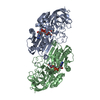
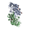
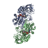
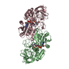
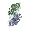
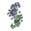
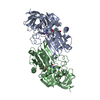
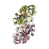
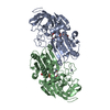
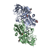
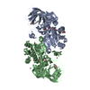
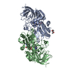
- PDB-1ee2: THE STRUCTURE OF STEROID-ACTIVE ALCOHOL DEHYDROGENASE AT 1.54 A R... -
+ Open data

- Basic information
| Entry | Database: PDB / ID: 1ee2 | ||||||
|---|---|---|---|---|---|---|---|
| Title | THE STRUCTURE OF STEROID-ACTIVE ALCOHOL DEHYDROGENASE AT 1.54 A RESOLUTION | ||||||
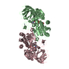
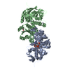
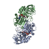
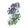
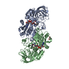
 Components Components | ALCOHOL DEHYDROGENASE | ||||||
 Keywords Keywords | OXIDOREDUCTASE / DEHYDROGENASE / ALCOHOL / NICOTINAMIDE COENZYME / STEROID BINDING | ||||||
| Function / homology |  Function and homology information Function and homology informationall-trans-retinol dehydrogenase (NAD+) activity / alcohol dehydrogenase / retinoic acid metabolic process / retinol metabolic process / zinc ion binding / cytosol Similarity search - Function | ||||||
| Biological species |  | ||||||
| Method |  X-RAY DIFFRACTION / SYNCHROTRON / Resolution: 1.54 Å X-RAY DIFFRACTION / SYNCHROTRON / Resolution: 1.54 Å | ||||||
 Authors Authors | Adolph, H.W. | ||||||
 Citation Citation | Journal: Biochemistry / Year: 2000 Title: Structural basis for substrate specificity differences of horse liver alcohol dehydrogenase isozymes. Authors: Adolph, H.W. / Zwart, P. / Meijers, R. / Hubatsch, I. / Kiefer, M. / Lamzin, V. / Cedergren-Zeppezauer, E. #1: Journal: Eur.J.Biochem. / Year: 1991Title: Substrate Specificity and Stereoselectivity of Horse Liver Alcohol Dehydrogenase. Kinetic Evaluation of Binding and Activation Parameters Controlling the Catalitic Cycles of Unbranched, ...Title: Substrate Specificity and Stereoselectivity of Horse Liver Alcohol Dehydrogenase. Kinetic Evaluation of Binding and Activation Parameters Controlling the Catalitic Cycles of Unbranched, Acyclic Secondary Alcohols and Ketones as Substrates of the Native and Active-Site-Specific Co(II)-Substituted Enzyme Authors: Adolph, H.W. / Maurer, P. / Scheinder-Bernlohr, H. / Sartorius, C. / Zeppezauer, M. #2: Journal: J.CHROMATOGR.,A / Year: 1995Title: Preparation and Characterization of Isozymes and Isoforms of Horse Liver Alcohol Dehydrogenase Authors: Hubatsch, I. / Maurer, P. / Engel, D. / Adolph, H.W. #3: Journal: Biochemistry / Year: 1997Title: Electrostatic Effects in the Kinetics of Coenzyme Binding to Isozymes of Alcohol Dehydrogenase from Horse Liver Authors: Adolph, H.W. / Kiefer, M. / Cedergren-Zeppezauer, E. | ||||||
| History |
|
- Structure visualization
| Structure viewer | Molecule: MolmilJmol/JSmol |
|---|
- Downloads & links
-Download
| PDBx/mmCIF format | 1ee2.cif.gz | 185.2 KB | Display | PDBx/mmCIF format |
|---|---|---|---|---|
| PDB format | pdb1ee2.ent.gz | 143.8 KB | Display | PDB format |
| PDBx/mmJSON format | 1ee2.json.gz | Tree view | PDBx/mmJSON format | |
| Others |  Other downloads Other downloads |
-Validation report
| Arichive directory | https://data.pdbj.org/pub/pdb/validation_reports/ee/1ee2ftp://data.pdbj.org/pub/pdb/validation_reports/ee/1ee2 | HTTPS FTP |
|---|
-Related structure data
| Related structure data | |
|---|---|
| Similar structure data |


-Links
 PDBj
PDBj

- Assembly
| Deposited unit | 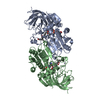
| ||||||||
|---|---|---|---|---|---|---|---|---|---|
| 1 |
| ||||||||
| Unit cell |
|
-Components
| #1: Protein | Mass: 39541.090 Da / Num. of mol.: 2 / Source method: isolated from a natural source / Source: (natural) #2: Chemical | ChemComp-ZN /   Mass: 65.409 Da / Num. of mol.: 4 / Source method: obtained synthetically / Formula: Zn Mass: 65.409 Da / Num. of mol.: 4 / Source method: obtained synthetically / Formula: Zn#3: Chemical |   Mass: 663.425 Da / Num. of mol.: 2 / Source method: obtained synthetically / Formula: C21H27N7O14P2 / Comment: NAD*YM Mass: 663.425 Da / Num. of mol.: 2 / Source method: obtained synthetically / Formula: C21H27N7O14P2 / Comment: NAD*YM#4: Chemical | 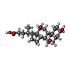  Mass: 408.571 Da / Num. of mol.: 2 / Source method: obtained synthetically / Formula: C24H40O5 Mass: 408.571 Da / Num. of mol.: 2 / Source method: obtained synthetically / Formula: C24H40O5#5: Water | ChemComp-HOH / |  Mass: 18.015 Da / Num. of mol.: 1010 / Source method: isolated from a natural source / Formula: H2O Mass: 18.015 Da / Num. of mol.: 1010 / Source method: isolated from a natural source / Formula: H2O |
|---|
-Experimental details
-Experiment
| Experiment | Method: X-RAY DIFFRACTION / Number of used crystals: 1 |
|---|
- Sample preparation
| Crystal | Density Matthews: 2.3 Å3/Da / Density % sol: 46.46 % | ||||||||||||||||||||||||||||||||||||
|---|---|---|---|---|---|---|---|---|---|---|---|---|---|---|---|---|---|---|---|---|---|---|---|---|---|---|---|---|---|---|---|---|---|---|---|---|---|
| Crystal grow | Temperature: 277 K / Method: vapor diffusion, sitting drop / pH: 7 Details: PEG400, PEG8000, TES/KOH, NAD+, Cholic Acid, pH 7, VAPOR DIFFUSION, SITTING DROP, temperature 277K | ||||||||||||||||||||||||||||||||||||
| Crystal grow | *PLUS Details: used micro bridge | ||||||||||||||||||||||||||||||||||||
| Components of the solutions | *PLUS
|
-Data collection
| Diffraction | Mean temperature: 100 K |
|---|---|
| Diffraction source | Source: SYNCHROTRON / Site: EMBL/DESY, HAMBURG  / Beamline: BW7B / Wavelength: 0.8373 / Beamline: BW7B / Wavelength: 0.8373 |
| Detector | Type: MARRESEARCH / Detector: IMAGE PLATE / Date: Nov 23, 1998 |
| Radiation | Protocol: SINGLE WAVELENGTH / Monochromatic (M) / Laue (L): M / Scattering type: x-ray |
| Radiation wavelength | Wavelength: 0.8373 Å / Relative weight: 1 |
| Reflection | Resolution: 1.54→20 Å / Num. all: 103854 / Num. obs: 103854 / % possible obs: 98 % / Observed criterion σ(F): 0 / Observed criterion σ(I): 0 / Redundancy: 4.16 % / Biso Wilson estimate: 14.1 Å2 / Rmerge(I) obs: 0.106 / Net I/σ(I): 10.9 |
| Reflection shell | Resolution: 1.54→1.57 Å / Redundancy: 1.8 % / Rmerge(I) obs: 0.25 / % possible all: 97.2 |
| Reflection | *PLUS % possible obs: 98.3 % |
| Reflection shell | *PLUS % possible obs: 97.2 % / Mean I/σ(I) obs: 4.5 |
- Processing
| Software |
| ||||||||||||||||||||
|---|---|---|---|---|---|---|---|---|---|---|---|---|---|---|---|---|---|---|---|---|---|
| Refinement | Resolution: 1.54→20 Å / σ(F): 0 / σ(I): 0 / Stereochemistry target values: Eng & Huber
| ||||||||||||||||||||
| Refinement step | Cycle: LAST / Resolution: 1.54→20 Å
| ||||||||||||||||||||
| Refine LS restraints |
| ||||||||||||||||||||
| Software | *PLUS Name: REFMAC / Classification: refinement | ||||||||||||||||||||
| Refinement | *PLUS Lowest resolution: 20 Å / σ(F): 0 / % reflection Rfree: 2 % / Rfactor obs: 0.145 | ||||||||||||||||||||
| Solvent computation | *PLUS | ||||||||||||||||||||
| Displacement parameters | *PLUS | ||||||||||||||||||||
| Refine LS restraints | *PLUS
|
