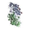
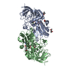
 Movie
Movie Controller

[English] 日本語
 Yorodumi
- PDB-1p0f: Crystal Structure of the Binary Complex: NADP(H)-Dependent Verteb... -
+ Open data

- Basic information
| Entry | Database: PDB / ID: 1p0f | ||||||
|---|---|---|---|---|---|---|---|
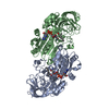
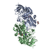
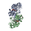
| Title | Crystal Structure of the Binary Complex: NADP(H)-Dependent Vertebrate Alcohol Dehydrogenase (ADH8) with the cofactor NADP | ||||||
 Components Components | NADP-dependent ALCOHOL DEHYDROGENASE | ||||||
 Keywords Keywords | OXIDOREDUCTASE / ADH TOPOLOGY / NADP(H)-DEPENDENT | ||||||
| Function / homology |  Function and homology information Function and homology informationalcohol dehydrogenase (NADP+) / all-trans-retinol dehydrogenase (NAD+) activity / alcohol dehydrogenase (NADP+) activity / retinoic acid metabolic process / retinol metabolic process / zinc ion binding / cytosol Similarity search - Function | ||||||
| Biological species | Rana perezi (Iberian green frog) | ||||||
| Method |  X-RAY DIFFRACTION / SYNCHROTRON / MOLECULAR REPLACEMENT / Resolution: 1.8 Å X-RAY DIFFRACTION / SYNCHROTRON / MOLECULAR REPLACEMENT / Resolution: 1.8 Å | ||||||
 Authors Authors | Rosell, A. / Valencia, E. / Pares, X. / Fita, I. / Farres, J. / Ochoa, W.F. | ||||||
 Citation Citation | Journal: J.Mol.Biol. / Year: 2003 Title: Crystal structure of the vertebrate NADP(H)-dependent alcohol dehydrogenase (ADH8) Authors: Rosell, A. / Valencia, E. / Pares, X. / Fita, I. / Farres, J. / Ochoa, W.F. #1: Journal: To be published / Year: 2003Title: Crystallization and preliminary X-ray analysis of NADP(H)-dependent alcohol dehydrogenases from Saccharomyces cerevisiae and Rana perezi Authors: Valencia, E. / Rosell, A. / Larroy, C. / Farres, J. / Biosca, J.A. / Fita, I. / Pares, X. / Ochoa, W.F. | ||||||
| History |
|
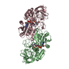
- Structure visualization
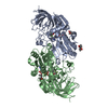
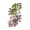
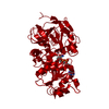
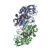
| Structure viewer | Molecule: MolmilJmol/JSmol |
|---|
- Downloads & links
-Download
| PDBx/mmCIF format | 1p0f.cif.gz | 160.2 KB | Display | PDBx/mmCIF format |
|---|---|---|---|---|
| PDB format | pdb1p0f.ent.gz | 123.9 KB | Display | PDB format |
| PDBx/mmJSON format | 1p0f.json.gz | Tree view | PDBx/mmJSON format | |
| Others |  Other downloads Other downloads |
-Validation report
| Arichive directory | https://data.pdbj.org/pub/pdb/validation_reports/p0/1p0fftp://data.pdbj.org/pub/pdb/validation_reports/p0/1p0f | HTTPS FTP |
|---|
-Related structure data


-Links
 PDBj
PDBj

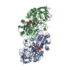
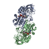
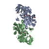
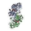
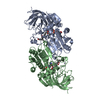
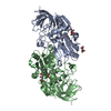
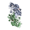
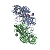
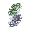
- Assembly
| Deposited unit | 
| ||||||||
|---|---|---|---|---|---|---|---|---|---|
| 1 |
| ||||||||
| Unit cell |
|
-Components
| #1: Protein | Mass: 39243.652 Da / Num. of mol.: 2 Source method: isolated from a genetically manipulated source Source: (gene. exp.) Rana perezi (Iberian green frog) / Gene: ADH / Plasmid: pGEX4T-2 / Species (production host): Escherichia coli / Production host:  #2: Chemical | ChemComp-ZN /   Mass: 65.409 Da / Num. of mol.: 4 / Source method: obtained synthetically / Formula: Zn Mass: 65.409 Da / Num. of mol.: 4 / Source method: obtained synthetically / Formula: Zn#3: Chemical |   Mass: 743.405 Da / Num. of mol.: 2 / Source method: obtained synthetically / Formula: C21H28N7O17P3 Mass: 743.405 Da / Num. of mol.: 2 / Source method: obtained synthetically / Formula: C21H28N7O17P3#4: Chemical |   Mass: 92.094 Da / Num. of mol.: 2 / Source method: obtained synthetically / Formula: C3H8O3 Mass: 92.094 Da / Num. of mol.: 2 / Source method: obtained synthetically / Formula: C3H8O3#5: Water | ChemComp-HOH / |  Mass: 18.015 Da / Num. of mol.: 397 / Source method: isolated from a natural source / Formula: H2O Mass: 18.015 Da / Num. of mol.: 397 / Source method: isolated from a natural source / Formula: H2O |
|---|
-Experimental details
-Experiment
| Experiment | Method: X-RAY DIFFRACTION |
|---|
- Sample preparation
| Crystal | Density Matthews: 2.46 Å3/Da / Density % sol: 49.58 % | |||||||||||||||
|---|---|---|---|---|---|---|---|---|---|---|---|---|---|---|---|---|
| Crystal grow | Temperature: 277 K / Method: vapor diffusion, hanging drop / pH: 8 Details: PEG 4000, LiSO4, pH 8.0, VAPOR DIFFUSION, HANGING DROP, temperature 277.0K | |||||||||||||||
| Crystal grow | *PLUS Temperature: 293 KDetails: Valencia, E., (2003) Acta Crystallogr., Sect.D, 59, 334. | |||||||||||||||
| Components of the solutions | *PLUS
|
-Data collection
| Diffraction | Mean temperature: 100 K |
|---|---|
| Diffraction source | Source: SYNCHROTRON / Site: ESRF  / Beamline: ID14-4 / Wavelength: 0.953 Å / Beamline: ID14-4 / Wavelength: 0.953 Å |
| Detector | Type: ADSC QUANTUM 4 / Detector: CCD / Date: Mar 8, 2002 |
| Radiation | Protocol: SINGLE WAVELENGTH / Monochromatic (M) / Laue (L): M / Scattering type: x-ray |
| Radiation wavelength | Wavelength: 0.953 Å / Relative weight: 1 |
| Reflection | Resolution: 1.8→30 Å / % possible obs: 96.1 % / Observed criterion σ(F): 0 / Observed criterion σ(I): 0 |
| Reflection | *PLUS Lowest resolution: 30 Å / Num. obs: 140960 / Num. measured all: 254342 / Rmerge(I) obs: 0.048 |
| Reflection shell | *PLUS Highest resolution: 1.9 Å / % possible obs: 74 % / Rmerge(I) obs: 0.331 / Mean I/σ(I) obs: 2 |
- Processing
| Software |
| ||||||||||||||||||||||||||||||||||||||||||||||||||||||||||||||||||||||||||||||||
|---|---|---|---|---|---|---|---|---|---|---|---|---|---|---|---|---|---|---|---|---|---|---|---|---|---|---|---|---|---|---|---|---|---|---|---|---|---|---|---|---|---|---|---|---|---|---|---|---|---|---|---|---|---|---|---|---|---|---|---|---|---|---|---|---|---|---|---|---|---|---|---|---|---|---|---|---|---|---|---|---|---|
| Refinement | Method to determine structure: MOLECULAR REPLACEMENT / Resolution: 1.8→30 Å / Cross valid method: THROUGHOUT / σ(F): 0 / σ(I): 0
| ||||||||||||||||||||||||||||||||||||||||||||||||||||||||||||||||||||||||||||||||
| Displacement parameters |
| ||||||||||||||||||||||||||||||||||||||||||||||||||||||||||||||||||||||||||||||||
| Refinement step | Cycle: LAST / Resolution: 1.8→30 Å
| ||||||||||||||||||||||||||||||||||||||||||||||||||||||||||||||||||||||||||||||||
| Refine LS restraints |
| ||||||||||||||||||||||||||||||||||||||||||||||||||||||||||||||||||||||||||||||||
| Xplor file |
| ||||||||||||||||||||||||||||||||||||||||||||||||||||||||||||||||||||||||||||||||
| Refinement | *PLUS Lowest resolution: 30 Å / % reflection Rfree: 5 % / Rfactor Rfree: 0.223 / Rfactor Rwork: 0.199 | ||||||||||||||||||||||||||||||||||||||||||||||||||||||||||||||||||||||||||||||||
| Solvent computation | *PLUS | ||||||||||||||||||||||||||||||||||||||||||||||||||||||||||||||||||||||||||||||||
| Displacement parameters | *PLUS | ||||||||||||||||||||||||||||||||||||||||||||||||||||||||||||||||||||||||||||||||
| Refine LS restraints | *PLUS
|
