 Movie
Movie Controller


+ Open data

- Basic information
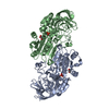
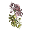
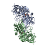
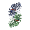
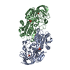
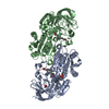
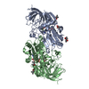
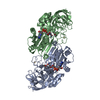
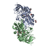
| Entry | Database: PDB / ID: 1ju9 | ||||||
|---|---|---|---|---|---|---|---|
| Title | HORSE LIVER ALCOHOL DEHYDROGENASE VAL292SER MUTANT | ||||||
 Components Components | ALCOHOL DEHYDROGENASE | ||||||
 Keywords Keywords | OXIDOREDUCTASE / DEHYDROGENASE / ALCOHOL / NICOTINAMIDE COENZYME / MUTANT | ||||||
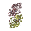
| Function / homology |  Function and homology information Function and homology informationall-trans-retinol dehydrogenase (NAD+) activity / alcohol dehydrogenase / retinoic acid metabolic process / retinol metabolic process / zinc ion binding / cytosol Similarity search - Function | ||||||
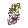
| Biological species |  | ||||||
| Method |  X-RAY DIFFRACTION / MOLECULAR REPLACEMENT / Resolution: 2 Å X-RAY DIFFRACTION / MOLECULAR REPLACEMENT / Resolution: 2 Å | ||||||
 Authors Authors | Rubach, J.K. / Ramaswamy, S. / Plapp, B.V. | ||||||
 Citation Citation | Journal: Biochemistry / Year: 2001 Title: Contributions of valine-292 in the nicotinamide binding site of liver alcohol dehydrogenase and dynamics to catalysis. Authors: Rubach, J.K. / Ramaswamy, S. / Plapp, B.V. #1: Journal: Biochemistry / Year: 1999Title: SUBSTITUTIONS IN THE FLEXIBLE LOOP OF HORSE LIVER ALCOHOL DEHYDROGENASE HINDER THE CONFORMATIONAL CHANGE AND UNMASK HYDROGEN TRANSFER Authors: Ramaswamy, S. / Park, D.H. / Plapp, B.V. #2: Journal: J.Biol.Chem. / Year: 1982Title: Binding of Substrate in a Ternary Complex of Horse Liver Alcohol Dehydrogenase Authors: Eklund, H. / Plapp, B.V. / Samama, J.-P. / Branden, C.-I. #3: Journal: J.Mol.Biol. / Year: 1981Title: Structure of a Triclinic Ternary Complex of Horse Liver Alcohol Dehydrogenase at 2.9 Angstroms Resolution Authors: Eklund, H. / Samama, J.-P. / Wallen, L. / Branden, C.-I. / Akeson, A. / A Jones, T. #4: Journal: J.Mol.Biol. / Year: 1976Title: Three-Dimensional Structure of Horse Liver Alcohol Dehydrogenase at 2.4 Angstroms Resolution Authors: Eklund, H. / Nordstrom, B. / Zeppezauer, E. / Soderlund, G. / Ohlsson, I. / Boiwe, T. / Soderberg, B.-O. / Tapia, O. / Branden, C.-I. / Akeson, A. | ||||||
| History |
| ||||||
| Remark 11 | THE RECOMBINANT ENZYME WAS CRYSTALLIZED IN HANGING DROPS CONTAINING 50 MM AMMMONIUM ...THE RECOMBINANT ENZYME WAS CRYSTALLIZED IN HANGING DROPS CONTAINING 50 MM AMMMONIUM N[TRIS(HYDROXYMETHYL)METHYL]- AMINOMETHANESULFONATE BUFFER, PH 7.0, AT 5 DEG C, WITH 10.6 MM NAD+ AND 20 MM 2,3,4,5,6-PENTAFLUOROBENZYL ALCOHOL AND 9% MPD OVER A RESERVOIR WITH 18 % MPD. HOWEVER, ONLY THE ADP PORTION OF NAD+ WAS VISIBLE IN THE ELECTRON DENSITY MAPS. THE COORDINATES ONLY INCLUDE THE ADP. |
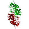
- Structure visualization
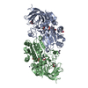
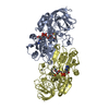
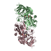
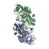
| Structure viewer | Molecule: MolmilJmol/JSmol |
|---|
- Downloads & links
-Download
| PDBx/mmCIF format | 1ju9.cif.gz | 154.2 KB | Display | PDBx/mmCIF format |
|---|---|---|---|---|
| PDB format | pdb1ju9.ent.gz | 120 KB | Display | PDB format |
| PDBx/mmJSON format | 1ju9.json.gz | Tree view | PDBx/mmJSON format | |
| Others |  Other downloads Other downloads |
-Validation report
| Arichive directory | https://data.pdbj.org/pub/pdb/validation_reports/ju/1ju9ftp://data.pdbj.org/pub/pdb/validation_reports/ju/1ju9 | HTTPS FTP |
|---|
-Related structure data
| Related structure data | 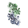 1hldS S: Starting model for refinement |
|---|---|
| Similar structure data |


-Links
 PDBj
PDBj

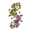
- Assembly
| Deposited unit | 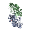
| ||||||||
|---|---|---|---|---|---|---|---|---|---|
| 1 |
| ||||||||
| Unit cell |
|
-Components
| #1: Protein | Mass: 39841.223 Da / Num. of mol.: 2 / Mutation: V292S Source method: isolated from a genetically manipulated source Source: (gene. exp.)  #2: Chemical | ChemComp-ZN /   Mass: 65.409 Da / Num. of mol.: 4 / Source method: obtained synthetically / Formula: Zn Mass: 65.409 Da / Num. of mol.: 4 / Source method: obtained synthetically / Formula: Zn#3: Chemical |   Mass: 663.425 Da / Num. of mol.: 2 / Source method: obtained synthetically / Formula: C21H27N7O14P2 / Comment: NAD*YM Mass: 663.425 Da / Num. of mol.: 2 / Source method: obtained synthetically / Formula: C21H27N7O14P2 / Comment: NAD*YM#4: Water | ChemComp-HOH / |  Mass: 18.015 Da / Num. of mol.: 164 / Source method: isolated from a natural source / Formula: H2O Mass: 18.015 Da / Num. of mol.: 164 / Source method: isolated from a natural source / Formula: H2O |
|---|
-Experimental details
-Experiment
| Experiment | Method: X-RAY DIFFRACTION / Number of used crystals: 1 |
|---|
- Sample preparation
| Crystal | Density Matthews: 2.32 Å3/Da / Density % sol: 46.89 % | ||||||||||||||||||||||||||||||||||||||||||
|---|---|---|---|---|---|---|---|---|---|---|---|---|---|---|---|---|---|---|---|---|---|---|---|---|---|---|---|---|---|---|---|---|---|---|---|---|---|---|---|---|---|---|---|
| Crystal grow | Temperature: 277 K / Method: vapor diffusion, hanging drop / pH: 7 Details: MPD, pH 7.0, VAPOR DIFFUSION, HANGING DROP, temperature 277K | ||||||||||||||||||||||||||||||||||||||||||
| Crystal grow | *PLUS Temperature: 5 ℃ | ||||||||||||||||||||||||||||||||||||||||||
| Components of the solutions | *PLUS
|
-Data collection
| Diffraction | Mean temperature: 100 K |
|---|---|
| Diffraction source | Source: ROTATING ANODE / Type: RIGAKU / Wavelength: 1.5418 Å |
| Detector | Type: RIGAKU RAXIS IV++ / Detector: IMAGE PLATE / Date: Sep 18, 2000 / Details: CONFOCAL |
| Radiation | Protocol: SINGLE WAVELENGTH / Monochromatic (M) / Laue (L): M / Scattering type: x-ray |
| Radiation wavelength | Wavelength: 1.5418 Å / Relative weight: 1 |
| Reflection | Resolution: 2→28.63 Å / Num. all: 41422 / Num. obs: 41422 / % possible obs: 90.7 % / Observed criterion σ(F): 0 / Observed criterion σ(I): 0 / Redundancy: 2 % / Biso Wilson estimate: 26.9 Å2 / Rmerge(I) obs: 0.037 / Rsym value: 0.037 / Net I/σ(I): 11.4 |
| Reflection shell | Resolution: 2.01→2.07 Å / Redundancy: 1.6 % / Rmerge(I) obs: 0.065 / Mean I/σ(I) obs: 8.9 / Rsym value: 0.065 / % possible all: 100 |
| Reflection | *PLUS Lowest resolution: 20 Å / Num. obs: 41073 / % possible obs: 90.8 % / Num. measured all: 82843 |
| Reflection shell | *PLUS % possible obs: 84.9 % |
- Processing
| Software |
| |||||||||||||||||||||||||||||||||||||||||||||||||||||||||||||||||||||||||||||||||||||||||||||||||||||||||
|---|---|---|---|---|---|---|---|---|---|---|---|---|---|---|---|---|---|---|---|---|---|---|---|---|---|---|---|---|---|---|---|---|---|---|---|---|---|---|---|---|---|---|---|---|---|---|---|---|---|---|---|---|---|---|---|---|---|---|---|---|---|---|---|---|---|---|---|---|---|---|---|---|---|---|---|---|---|---|---|---|---|---|---|---|---|---|---|---|---|---|---|---|---|---|---|---|---|---|---|---|---|---|---|---|---|---|
| Refinement | Method to determine structure: MOLECULAR REPLACEMENT Starting model: PDB ENTRY 1HLD Resolution: 2→20 Å / SU B: 5.07 / SU ML: 0.143 / Cross valid method: THROUGHOUT / σ(F): 0 / σ(I): 0 / ESU R: 0.25 / ESU R Free: 0.19 / Stereochemistry target values: refmac 5.0 mon_lib
| |||||||||||||||||||||||||||||||||||||||||||||||||||||||||||||||||||||||||||||||||||||||||||||||||||||||||
| Displacement parameters | Biso mean: 28.007 Å2
| |||||||||||||||||||||||||||||||||||||||||||||||||||||||||||||||||||||||||||||||||||||||||||||||||||||||||
| Refinement step | Cycle: LAST / Resolution: 2→20 Å
| |||||||||||||||||||||||||||||||||||||||||||||||||||||||||||||||||||||||||||||||||||||||||||||||||||||||||
| Refine LS restraints |
| |||||||||||||||||||||||||||||||||||||||||||||||||||||||||||||||||||||||||||||||||||||||||||||||||||||||||
| LS refinement shell | Resolution: 2→2.051 Å / Total num. of bins used: 20 /
| |||||||||||||||||||||||||||||||||||||||||||||||||||||||||||||||||||||||||||||||||||||||||||||||||||||||||
| Software | *PLUS Name: REFMAC / Version: 5 / Classification: refinement | |||||||||||||||||||||||||||||||||||||||||||||||||||||||||||||||||||||||||||||||||||||||||||||||||||||||||
| Refinement | *PLUS σ(F): 0 / % reflection Rfree: 1.5 % | |||||||||||||||||||||||||||||||||||||||||||||||||||||||||||||||||||||||||||||||||||||||||||||||||||||||||
| Solvent computation | *PLUS | |||||||||||||||||||||||||||||||||||||||||||||||||||||||||||||||||||||||||||||||||||||||||||||||||||||||||
| Displacement parameters | *PLUS |
