 Movie
Movie Controller

[English] 日本語
 Yorodumi
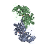
- PDB-6oa7: Horse liver L57F alcohol dehydrogenase E complexed with NAD and t... -
+ Open data

- Basic information
| Entry | Database: PDB / ID: 6oa7 | |||||||||
|---|---|---|---|---|---|---|---|---|---|---|
| Title | Horse liver L57F alcohol dehydrogenase E complexed with NAD and trifluoroethanol | |||||||||
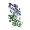
 Components Components | Alcohol dehydrogenase E chain | |||||||||
 Keywords Keywords | OXIDOREDUCTASE / alcohol dehydrogenase / NAD / 2 / 2-trifluoroethanol / Leu57 to Phe57 substitution / Horse liver E enzyme | |||||||||
| Function / homology |  Function and homology information Function and homology informationall-trans-retinol dehydrogenase (NAD+) activity / alcohol dehydrogenase / retinoic acid metabolic process / retinol metabolic process / zinc ion binding / cytosol Similarity search - Function | |||||||||
| Biological species |  | |||||||||
| Method |  X-RAY DIFFRACTION / SYNCHROTRON / MOLECULAR REPLACEMENT / Resolution: 1.1 Å X-RAY DIFFRACTION / SYNCHROTRON / MOLECULAR REPLACEMENT / Resolution: 1.1 Å | |||||||||
 Authors Authors | Plapp, B.V. | |||||||||
| Funding support |  United States, 2items United States, 2items
| |||||||||
 Citation Citation | Journal: Biochemistry / Year: 2020 Title: Substitutions of Amino Acid Residues in the Substrate Binding Site of Horse Liver Alcohol Dehydrogenase Have Small Effects on the Structures but Significantly Affect Catalysis of Hydrogen Transfer. Authors: Kim, K. / Plapp, B.V. #1: Journal: Biochemistry / Year: 1993 Title: Unmasking of hydrogen tunneling in the horse liver alcohol dehydrogenase reaction by site-directed mutagenesis. Authors: Bahnson, B.J. / Park, D.H. / Kim, K. / Plapp, B.V. / Klinman, J.P. #2: Journal: Biochemistry / Year: 2012Title: Atomic-resolution structures of horse liver alcohol dehydrogenase with NAD(+) and fluoroalcohols define strained Michaelis complexes. Authors: Plapp, B.V. / Ramaswamy, S. #3: Journal: Isotope Effects in Chemistry and Biology / Year: 2006Title: Catalysis by Alcohol Dehydrogenases Authors: Plapp, B.V. #4: Journal: Chem. Biol. Interact. / Year: 2017Title: Inversion of substrate stereoselectivity of horse liver alcohol dehydrogenase by substitutions of Ser-48 and Phe-93. Authors: Kim, K. / Plapp, B.V. | |||||||||
| History |
|
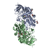
- Structure visualization
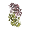
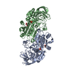
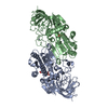
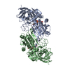
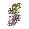
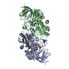
| Structure viewer | Molecule: MolmilJmol/JSmol |
|---|
- Downloads & links
-Download
| PDBx/mmCIF format | 6oa7.cif.gz | 358.5 KB | Display | PDBx/mmCIF format |
|---|---|---|---|---|
| PDB format | pdb6oa7.ent.gz | 290.5 KB | Display | PDB format |
| PDBx/mmJSON format | 6oa7.json.gz | Tree view | PDBx/mmJSON format | |
| Others |  Other downloads Other downloads |
-Validation report
| Arichive directory | https://data.pdbj.org/pub/pdb/validation_reports/oa/6oa7ftp://data.pdbj.org/pub/pdb/validation_reports/oa/6oa7 | HTTPS FTP |
|---|
-Related structure data
| Related structure data | 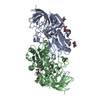 6o91SC 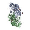 6owmC  6owpC S: Starting model for refinement C: citing same article ( |
|---|---|
| Similar structure data | |
| Experimental dataset #1 | Data reference: 10.15785/SBGRID/653 / Data set type: diffraction image data |


-Links
 PDBj
PDBj

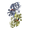
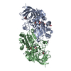
- Assembly
| Deposited unit | 
| ||||||||
|---|---|---|---|---|---|---|---|---|---|
| 1 |
| ||||||||
| Unit cell |
|
-Components
-Protein , 1 types, 2 molecules AB
| #1: Protein | Mass: 39887.289 Da / Num. of mol.: 2 / Mutation: L57F Source method: isolated from a genetically manipulated source Source: (gene. exp.)  |
|---|
-Non-polymers , 5 types, 1091 molecules 
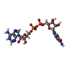
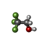


| #2: Chemical | ChemComp-ZN /  Mass: 65.409 Da / Num. of mol.: 4 / Source method: obtained synthetically / Formula: Zn Mass: 65.409 Da / Num. of mol.: 4 / Source method: obtained synthetically / Formula: Zn#3: Chemical |  Mass: 663.425 Da / Num. of mol.: 2 / Source method: obtained synthetically / Formula: C21H27N7O14P2 Mass: 663.425 Da / Num. of mol.: 2 / Source method: obtained synthetically / Formula: C21H27N7O14P2#4: Chemical |  Mass: 100.040 Da / Num. of mol.: 2 / Source method: obtained synthetically / Formula: C2H3F3O Mass: 100.040 Da / Num. of mol.: 2 / Source method: obtained synthetically / Formula: C2H3F3O#5: Chemical | ChemComp-MRD / (  Mass: 118.174 Da / Num. of mol.: 5 / Source method: obtained synthetically / Formula: C6H14O2 / Comment: precipitant*YM Mass: 118.174 Da / Num. of mol.: 5 / Source method: obtained synthetically / Formula: C6H14O2 / Comment: precipitant*YM#6: Water | ChemComp-HOH / | Mass: 18.015 Da / Num. of mol.: 1078 / Source method: isolated from a natural source / Formula: H2O |
|---|
-Experimental details
-Experiment
| Experiment | Method: X-RAY DIFFRACTION / Number of used crystals: 1 |
|---|
- Sample preparation
| Crystal | Density Matthews: 2.4 Å3/Da / Density % sol: 48.86 % / Description: block |
|---|---|
| Crystal grow | Temperature: 278 K / Method: microdialysis / pH: 7 Details: 50 MM AMMONIUM N-[TRIS(HYDROXYMETHYL)METHYL]-2-AMINOETHANE SULFONATE, PH 6.7 (AT 25 C), 0.25 MM EDTA, 10 MG/ML PROTEIN, 1 MM NAD+, 100 MM 2,2,2-trifluoroethanol, 12 TO 25 % 2-METHYL-2,4-PENTANEDIOL, PH 7.0 |
-Data collection
| Diffraction | Mean temperature: 100 K / Serial crystal experiment: N | ||||||||||||||||||||||||||||||||||||||||||||||||||||||||||||||||||||||||||||||||||||||||||||||||||||||||||||||
|---|---|---|---|---|---|---|---|---|---|---|---|---|---|---|---|---|---|---|---|---|---|---|---|---|---|---|---|---|---|---|---|---|---|---|---|---|---|---|---|---|---|---|---|---|---|---|---|---|---|---|---|---|---|---|---|---|---|---|---|---|---|---|---|---|---|---|---|---|---|---|---|---|---|---|---|---|---|---|---|---|---|---|---|---|---|---|---|---|---|---|---|---|---|---|---|---|---|---|---|---|---|---|---|---|---|---|---|---|---|---|---|
| Diffraction source | Source: SYNCHROTRON / Site: APS / Beamline: 23-ID-B / Wavelength: 1.0332 Å | ||||||||||||||||||||||||||||||||||||||||||||||||||||||||||||||||||||||||||||||||||||||||||||||||||||||||||||||
| Detector | Type: MARMOSAIC 300 mm CCD / Detector: CCD / Date: Jun 16, 2007 / Details: ADJUSTABLE FOCUS K-B PAIR SIPLUS PT, RH COATINGS | ||||||||||||||||||||||||||||||||||||||||||||||||||||||||||||||||||||||||||||||||||||||||||||||||||||||||||||||
| Radiation | Monochromator: DOUBLE CRYSTAL CRYOCOOLED SI(111) / Protocol: SINGLE WAVELENGTH / Monochromatic (M) / Laue (L): M / Scattering type: x-ray | ||||||||||||||||||||||||||||||||||||||||||||||||||||||||||||||||||||||||||||||||||||||||||||||||||||||||||||||
| Radiation wavelength | Wavelength: 1.0332 Å / Relative weight: 1 | ||||||||||||||||||||||||||||||||||||||||||||||||||||||||||||||||||||||||||||||||||||||||||||||||||||||||||||||
| Reflection | Resolution: 1.1→19.43 Å / Num. obs: 267237 / % possible obs: 88.5 % / Redundancy: 5.35 % / Rmerge(I) obs: 0.055 / Rrim(I) all: 0.06 / Χ2: 1.17 / Net I/σ(I): 17.2 / Num. measured all: 1440609 / Scaling rejects: 10806 | ||||||||||||||||||||||||||||||||||||||||||||||||||||||||||||||||||||||||||||||||||||||||||||||||||||||||||||||
| Reflection shell | Diffraction-ID: 1
|
- Processing
| Software |
| |||||||||||||||||||||||||||||||||||||||||||||||||||||||||||||||||
|---|---|---|---|---|---|---|---|---|---|---|---|---|---|---|---|---|---|---|---|---|---|---|---|---|---|---|---|---|---|---|---|---|---|---|---|---|---|---|---|---|---|---|---|---|---|---|---|---|---|---|---|---|---|---|---|---|---|---|---|---|---|---|---|---|---|---|
| Refinement | Method to determine structure: MOLECULAR REPLACEMENT Starting model: 6o91 Resolution: 1.1→19.43 Å / Cor.coef. Fo:Fc: 0.982 / Cor.coef. Fo:Fc free: 0.979 / SU B: 0.607 / SU ML: 0.014 / Cross valid method: THROUGHOUT / σ(F): 0 / ESU R: 0.025 / ESU R Free: 0.025 / Stereochemistry target values: MAXIMUM LIKELIHOOD Details: HYDROGENS HAVE BEEN ADDED IN THE RIDING POSITIONS U VALUES : REFINED INDIVIDUALLY
| |||||||||||||||||||||||||||||||||||||||||||||||||||||||||||||||||
| Solvent computation | Ion probe radii: 0.8 Å / Shrinkage radii: 0.8 Å / VDW probe radii: 1.2 Å / Solvent model: MASK | |||||||||||||||||||||||||||||||||||||||||||||||||||||||||||||||||
| Displacement parameters | Biso max: 65.82 Å2 / Biso mean: 14.835 Å2 / Biso min: 7.65 Å2
| |||||||||||||||||||||||||||||||||||||||||||||||||||||||||||||||||
| Refinement step | Cycle: final / Resolution: 1.1→19.43 Å
| |||||||||||||||||||||||||||||||||||||||||||||||||||||||||||||||||
| Refine LS restraints |
| |||||||||||||||||||||||||||||||||||||||||||||||||||||||||||||||||
| LS refinement shell | Resolution: 1.1→1.128 Å / Rfactor Rfree error: 0 / Total num. of bins used: 20
|
