 Movie
Movie Controller

[English] 日本語
 Yorodumi
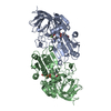
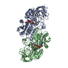
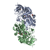
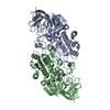
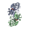
- PDB-1het: atomic X-ray structure of liver alcohol dehydrogenase containing ... -
+ Open data

- Basic information
| Entry | Database: PDB / ID: 1het | ||||||
|---|---|---|---|---|---|---|---|
| Title | atomic X-ray structure of liver alcohol dehydrogenase containing a hydroxide adduct to NADH | ||||||
 Components Components | ALCOHOL DEHYDROGENASE E CHAIN | ||||||
 Keywords Keywords | OXIDOREDUCTASE / OXIDOREDUCTASE(NAD(A)-CHOH(D)) | ||||||
| Function / homology |  Function and homology information Function and homology informationall-trans-retinol dehydrogenase (NAD+) activity / alcohol dehydrogenase / retinoic acid metabolic process / retinol metabolic process / zinc ion binding / cytosol Similarity search - Function | ||||||
| Biological species |  | ||||||
| Method |  X-RAY DIFFRACTION / SYNCHROTRON / MOLECULAR REPLACEMENT / Resolution: 1.15 Å X-RAY DIFFRACTION / SYNCHROTRON / MOLECULAR REPLACEMENT / Resolution: 1.15 Å | ||||||
 Authors Authors | Meijers, R. / Morris, R.J. / Adolph, H.W. / Merli, A. / Lamzin, V.S. / Cedergen-Zeppezauer, E.S. | ||||||
 Citation Citation | Journal: J.Biol.Chem. / Year: 2001 Title: On the Enzymatic Activation of Nadh Authors: Meijers, R. / Morris, R.J. / Adolph, H.W. / Merli, A. / Lamzin, V.S. / Cedergen-Zeppezauer, E.S. #1: Journal: Biochemistry / Year: 1999Title: Substitutions in a Flexible Loop of Horse Liver Alcohol Dehydrogenase Hinder the Conformational Change and Unmask Hydrogen Transfer Authors: Ramaswamy, S. / Park, D.H. / Plapp, B.V. #2: Journal: Biochemistry / Year: 1997 Title: Electrostatic Effects in the Kinetics of Coenzyme Binding to Isozymes of Alcohol Dehydrogenase from Horse Liver Authors: Adolph, H.W. / Kiefer, M. / Cedergren-Zeppezauer, E.S. #3: Journal: Acta Crystallogr.,Sect.D / Year: 1995Title: Refined Structure of Cu-Substituted Alcohol Dehydrogenase at 2.1 A Resolution Authors: Al-Karadaghi, S. / Cedergren-Zeppezauer, E.S. / Dauter, Z. / Wilson, K.S. #4: Journal: Acta Crystallogr.,Sect.D / Year: 1994Title: Refined Crystal Structure of Liver Alcohol Dehydrogenase-Nadh Complex at 1.8 Angstrom Resolution Authors: Al-Karadaghi, S. / Cedergren-Zeppezauer, E.S. / Petratos, K. / Hovmoeller, S. / Terry, H. / Dauter, Z. / Wilson, K.S. #5: Journal: J.Biol.Chem. / Year: 1986Title: Interdomain Motion in Liver Alcohol Dehydrogenase. {S}Tructural and Energetic Analysis of the Hinge Bending Mode Authors: Colonna-Cesari, F. / Perahia, D. / Karplus, M. / Eklund, H. / Branden, C.I. / Tapia, O. #6: Journal: Biochemistry / Year: 1984 Title: Crystallographic Investigations of Nicotinamide Adenine Dinucleotide Authors: Eklund, H. / Samama, J.-P. / Jones, T.A. #7: Journal: Proc.Natl.Acad.Sci.USA / Year: 1983 Title: Crystal Structures of the Active Site in Specifically Metal-Depleted and Cobalt-Substituted Horse Liver Alcohol Dehydrogenase Derivatives Authors: Schneider, G. / Eklund, H. / Cedergren-Zeppezauer, E. / Zeppezauer, M. #8: Journal: Biochemistry / Year: 1983 Title: Crystal-Structure Determination of Reduced Nicotinamide Adenine Dinucleotide Complex with Horse Liver Alcohol Dehydrogenase Maintained in its Apo Conformation by Zinc-Bound Imidazole Authors: Cedergren-Zeppezauer, E. #9: Journal: Biochemistry / Year: 1982 Title: Crystal Structure Determinations of Coenzyme Analogue and Substrate Complexes of Liver Alcohol Dehydrogenase: Binding of 1,4,5,6-Tetrahydronicotinamide Adenine Dinucleotide and Trans-4- ...Title: Crystal Structure Determinations of Coenzyme Analogue and Substrate Complexes of Liver Alcohol Dehydrogenase: Binding of 1,4,5,6-Tetrahydronicotinamide Adenine Dinucleotide and Trans-4-({N},{N}-Dimethylamino)Cinnamaldehyde to the Enzyme Authors: Cedergren-Zeppezauer, E. / Samama, J.-P. / Eklund, H. #10: Journal: J.Mol.Biol. / Year: 1976 Title: Three-Dimensional Structure of Horse Liver Alcohol Dehydrogenase at 2.4 Angstrom Resolution Authors: Eklund, H. / Nordstrom, B. / Zeppezauer, E. / Soderlund, G. / Ohlsson, I. / Boiwe, T. / Soderberg, B.-O. / Tapia, O. / Branden, C.-I. / Akeson, A. #11: Journal: Proc.Natl.Acad.Sci.USA / Year: 1973 Title: Structure of Liver Alcohol Dehydrogenase at 2.9 Angstrom Resolution Authors: Branden, C.-I. / Eklund, H. / Nordstrom, B. / Boiwe, T. / Soderlund, G. / Zeppezauer, E. / Ohlsson, I. / Akeson, A. | ||||||
| History |
| ||||||
| Remark 650 | HELIX DETERMINATION METHOD: AUTHOR PROVIDED. | ||||||
| Remark 700 | SHEET DETERMINATION METHOD: AUTHOR PROVIDED. |
- Structure visualization
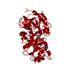
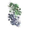
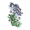
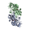
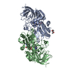
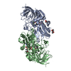
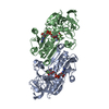
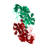
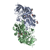
| Structure viewer | Molecule: MolmilJmol/JSmol |
|---|
- Downloads & links
-Download
| PDBx/mmCIF format | 1het.cif.gz | 377.6 KB | Display | PDBx/mmCIF format |
|---|---|---|---|---|
| PDB format | pdb1het.ent.gz | 309.6 KB | Display | PDB format |
| PDBx/mmJSON format | 1het.json.gz | Tree view | PDBx/mmJSON format | |
| Others |  Other downloads Other downloads |
-Validation report
| Arichive directory | https://data.pdbj.org/pub/pdb/validation_reports/he/1hetftp://data.pdbj.org/pub/pdb/validation_reports/he/1het | HTTPS FTP |
|---|
-Related structure data
| Related structure data | 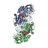 1heuC 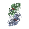 1hf3C 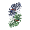 2ohxS C: citing same article ( S: Starting model for refinement |
|---|---|
| Similar structure data |


-Links
 PDBj
PDBj

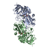
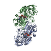
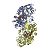
- Assembly
| Deposited unit | 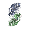
| ||||||||
|---|---|---|---|---|---|---|---|---|---|
| 1 |
| ||||||||
| Unit cell |
| ||||||||
| Noncrystallographic symmetry (NCS) | NCS oper: (Code: given Matrix: (0.12925, 0.98584, -0.10681), Vector: Details | BIOLOGICAL_UNIT: DIMERIC | |
-Components
| #1: Protein | Mass: 39853.273 Da / Num. of mol.: 2 / Source method: isolated from a natural source Details: ADDUCT BETWEEN NC6 OF NADH AND ZINC BOUND HYDROXIDE Source: (natural) #2: Chemical | ChemComp-ZN /   Mass: 65.409 Da / Num. of mol.: 4 / Source method: obtained synthetically / Formula: Zn Mass: 65.409 Da / Num. of mol.: 4 / Source method: obtained synthetically / Formula: Zn#3: Chemical |   Mass: 663.425 Da / Num. of mol.: 2 / Source method: obtained synthetically / Formula: C21H27N7O14P2 / Comment: NAD*YM Mass: 663.425 Da / Num. of mol.: 2 / Source method: obtained synthetically / Formula: C21H27N7O14P2 / Comment: NAD*YM#4: Chemical |   Mass: 118.174 Da / Num. of mol.: 3 / Source method: obtained synthetically / Formula: C6H14O2 / Comment: precipitant*YM Mass: 118.174 Da / Num. of mol.: 3 / Source method: obtained synthetically / Formula: C6H14O2 / Comment: precipitant*YM#5: Water | ChemComp-HOH / |  Mass: 18.015 Da / Num. of mol.: 1436 / Source method: isolated from a natural source / Formula: H2O Mass: 18.015 Da / Num. of mol.: 1436 / Source method: isolated from a natural source / Formula: H2OCompound details | ALCOHOL + NAD(+) = ALDEHYDE OR KETONE + NADH. REQUIRES ZINC FOR ITS ACTIVITY. DIMER OF IDENTICAL OR ...ALCOHOL + NAD(+) = ALDEHYDE OR KETONE + NADH. REQUIRES ZINC FOR ITS ACTIVITY. DIMER OF IDENTICAL OR NONIDENTIC | |
|---|
-Experimental details
-Experiment
| Experiment | Method: X-RAY DIFFRACTION / Number of used crystals: 1 |
|---|
- Sample preparation
| Crystal | Density Matthews: 2.42 Å3/Da / Density % sol: 49.2 % | ||||||||||||||||||||
|---|---|---|---|---|---|---|---|---|---|---|---|---|---|---|---|---|---|---|---|---|---|
| Crystal grow | pH: 8.2 / Details: pH 8.20 | ||||||||||||||||||||
| Crystal grow | *PLUS Method: unknown | ||||||||||||||||||||
| Components of the solutions | *PLUS
|
-Data collection
| Diffraction | Mean temperature: 120 K |
|---|---|
| Diffraction source | Source: SYNCHROTRON / Site: EMBL/DESY, HAMBURG  / Beamline: BW7B / Wavelength: 0.927 / Beamline: BW7B / Wavelength: 0.927 |
| Detector | Type: MARRESEARCH X-RAY RESEARCH / Detector: IMAGE PLATE |
| Radiation | Protocol: SINGLE WAVELENGTH / Monochromatic (M) / Laue (L): M / Scattering type: x-ray |
| Radiation wavelength | Wavelength: 0.927 Å / Relative weight: 1 |
| Reflection | Resolution: 1.1→20 Å / Num. obs: 267243 / % possible obs: 94.5 % / Redundancy: 2.6 % / Biso Wilson estimate: 9.4 Å2 / Rmerge(I) obs: 0.054 / Net I/σ(I): 13.1 |
| Reflection shell | Resolution: 1.1→1.12 Å / Rmerge(I) obs: 0.38 / Mean I/σ(I) obs: 2 / % possible all: 89.7 |
| Reflection | *PLUS Num. measured all: 684025 |
- Processing
| Software |
| |||||||||||||||||||||||||||||||||||||||||||||||||||||||||||||||
|---|---|---|---|---|---|---|---|---|---|---|---|---|---|---|---|---|---|---|---|---|---|---|---|---|---|---|---|---|---|---|---|---|---|---|---|---|---|---|---|---|---|---|---|---|---|---|---|---|---|---|---|---|---|---|---|---|---|---|---|---|---|---|---|---|
| Refinement | Method to determine structure: MOLECULAR REPLACEMENT Starting model: 2OHX Resolution: 1.15→20 Å / Cross valid method: THROUGHOUT UNTIL FINAL REFINEMENT ROUND / σ(F): 0 / ESU R: 0.03 Details: SHELX WAS USED IN PARALLEL TO REFINE OCCUPANCIES OF DISORDERED RESIDUES
| |||||||||||||||||||||||||||||||||||||||||||||||||||||||||||||||
| Displacement parameters | Biso mean: 19.8 Å2 | |||||||||||||||||||||||||||||||||||||||||||||||||||||||||||||||
| Refinement step | Cycle: LAST / Resolution: 1.15→20 Å
| |||||||||||||||||||||||||||||||||||||||||||||||||||||||||||||||
| Refine LS restraints |
| |||||||||||||||||||||||||||||||||||||||||||||||||||||||||||||||
| Software | *PLUS Name: REFMAC / Classification: refinement | |||||||||||||||||||||||||||||||||||||||||||||||||||||||||||||||
| Refinement | *PLUS Lowest resolution: 20 Å | |||||||||||||||||||||||||||||||||||||||||||||||||||||||||||||||
| Solvent computation | *PLUS | |||||||||||||||||||||||||||||||||||||||||||||||||||||||||||||||
| Displacement parameters | *PLUS |
