 Movie
Movie Controller

[English] 日本語
 Yorodumi
- PDB-1rfb: CRYSTAL STRUCTURE OF RECOMBINANT BOVINE INTERFERON-GAMMA AT 3.0 A... -
+ Open data

- Basic information
| Entry | Database: PDB / ID: 1rfb | ||||||
|---|---|---|---|---|---|---|---|
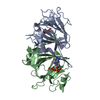
| Title | CRYSTAL STRUCTURE OF RECOMBINANT BOVINE INTERFERON-GAMMA AT 3.0 ANGSTROMS RESOLUTION | ||||||
 Components Components | INTERFERON-GAMMA | ||||||
 Keywords Keywords | GLYCOPROTEIN | ||||||
| Function / homology |  Function and homology information Function and homology informationInterferon gamma signaling / Regulation of IFNG signaling / IFNG signaling activates MAPKs / positive regulation of fructose 1,6-bisphosphate metabolic process / positive regulation of tumor necrosis factor (ligand) superfamily member 11 production / type II interferon receptor binding / : / positive regulation of vitamin D biosynthetic process / positive regulation of interleukin-23 production / type III interferon-mediated signaling pathway ...Interferon gamma signaling / Regulation of IFNG signaling / IFNG signaling activates MAPKs / positive regulation of fructose 1,6-bisphosphate metabolic process / positive regulation of tumor necrosis factor (ligand) superfamily member 11 production / type II interferon receptor binding / : / positive regulation of vitamin D biosynthetic process / positive regulation of interleukin-23 production / type III interferon-mediated signaling pathway / positive regulation of smooth muscle cell apoptotic process / negative regulation of interleukin-17 production / positive regulation of osteoclast differentiation / macrophage activation involved in immune response / positive regulation of membrane protein ectodomain proteolysis / positive regulation of neurogenesis / positive regulation of amyloid-beta formation / interleukin-2-mediated signaling pathway / Fc-gamma receptor signaling pathway involved in phagocytosis / regulation of insulin secretion / humoral immune response / macrophage differentiation / positive regulation of epithelial cell migration / type II interferon-mediated signaling pathway / cell surface receptor signaling pathway via JAK-STAT / extrinsic apoptotic signaling pathway / positive regulation of interleukin-12 production / positive regulation of B cell proliferation / positive regulation of chemokine production / positive regulation of autophagy / astrocyte activation / cytokine activity / negative regulation of smooth muscle cell proliferation / microglial cell activation / positive regulation of protein-containing complex assembly / positive regulation of T cell cytokine production / positive regulation of protein import into nucleus / positive regulation of interleukin-6 production / positive regulation of nitric oxide biosynthetic process / positive regulation of inflammatory response / defense response to virus / adaptive immune response / negative regulation of DNA-templated transcription / positive regulation of transcription by RNA polymerase II / : Similarity search - Function | ||||||
| Biological species |  | ||||||
| Method |  X-RAY DIFFRACTION / Resolution: 3 Å X-RAY DIFFRACTION / Resolution: 3 Å | ||||||
 Authors Authors | Samudzi, C.T. / Rubin, J.R. | ||||||
 Citation Citation | Journal: Acta Crystallogr.,Sect.D / Year: 1993 Title: Structure of recombinant bovine interferon-gamma at 3.0 A resolution. Authors: Samudzi, C.T. / Rubin, J.R. | ||||||
| History |
|
- Structure visualization
| Structure viewer | Molecule: MolmilJmol/JSmol |
|---|
- Downloads & links
-Download
| PDBx/mmCIF format | 1rfb.cif.gz | 60 KB | Display | PDBx/mmCIF format |
|---|---|---|---|---|
| PDB format | pdb1rfb.ent.gz | 46 KB | Display | PDB format |
| PDBx/mmJSON format | 1rfb.json.gz | Tree view | PDBx/mmJSON format | |
| Others |  Other downloads Other downloads |
-Validation report
| Arichive directory | https://data.pdbj.org/pub/pdb/validation_reports/rf/1rfbftp://data.pdbj.org/pub/pdb/validation_reports/rf/1rfb | HTTPS FTP |
|---|
-Related structure data
| Similar structure data |
|---|


-Links
 PDBj
PDBj


- Assembly
| Deposited unit | 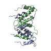
| ||||||||
|---|---|---|---|---|---|---|---|---|---|
| 1 |
| ||||||||
| Unit cell |
| ||||||||
| Atom site foot note | 1: SER B 19 - PRO B 20 OMEGA =279.70 PEPTIDE BOND DEVIATES SIGNIFICANTLY FROM TRANS CONFORMATION 2: GLY B 26 - PRO B 27 OMEGA =290.90 PEPTIDE BOND DEVIATES SIGNIFICANTLY FROM TRANS CONFORMATION |
-Components
| #1: Protein | Mass: 14055.080 Da / Num. of mol.: 2 Source method: isolated from a genetically manipulated source Source: (gene. exp.) #2: Water | ChemComp-HOH / |  Mass: 18.015 Da / Num. of mol.: 43 / Source method: isolated from a natural source / Formula: H2O Mass: 18.015 Da / Num. of mol.: 43 / Source method: isolated from a natural source / Formula: H2O |
|---|
-Experimental details
-Experiment
| Experiment | Method: X-RAY DIFFRACTION |
|---|
- Sample preparation
| Crystal | Density Matthews: 2.6 Å3/Da / Density % sol: 52.63 % | |||||||||||||||||||||||||
|---|---|---|---|---|---|---|---|---|---|---|---|---|---|---|---|---|---|---|---|---|---|---|---|---|---|---|
| Crystal grow | *PLUS Temperature: 17 ℃ / pH: 6 / Method: vapor diffusion | |||||||||||||||||||||||||
| Components of the solutions | *PLUS
|
-Data collection
| Radiation | Scattering type: x-ray |
|---|---|
| Radiation wavelength | Relative weight: 1 |
| Reflection | *PLUS Highest resolution: 2.66 Å / Num. obs: 7891 / Num. measured all: 19800 |
- Processing
| Software |
| ||||||||||||||||||||||||||||||||||||||||||||||||||||||||||||
|---|---|---|---|---|---|---|---|---|---|---|---|---|---|---|---|---|---|---|---|---|---|---|---|---|---|---|---|---|---|---|---|---|---|---|---|---|---|---|---|---|---|---|---|---|---|---|---|---|---|---|---|---|---|---|---|---|---|---|---|---|---|
| Refinement | Rfactor Rwork: 0.192 / Rfactor obs: 0.192 / Highest resolution: 3 Å | ||||||||||||||||||||||||||||||||||||||||||||||||||||||||||||
| Refinement step | Cycle: LAST / Highest resolution: 3 Å
| ||||||||||||||||||||||||||||||||||||||||||||||||||||||||||||
| Refine LS restraints |
| ||||||||||||||||||||||||||||||||||||||||||||||||||||||||||||
| Software | *PLUS Name: PROLSQ / Classification: refinement | ||||||||||||||||||||||||||||||||||||||||||||||||||||||||||||
| Refinement | *PLUS Highest resolution: 3 Å / Lowest resolution: 8 Å / Num. reflection obs: 2744 / σ(I): 2 / Rfactor obs: 0.192 | ||||||||||||||||||||||||||||||||||||||||||||||||||||||||||||
| Solvent computation | *PLUS | ||||||||||||||||||||||||||||||||||||||||||||||||||||||||||||
| Displacement parameters | *PLUS | ||||||||||||||||||||||||||||||||||||||||||||||||||||||||||||
| Refine LS restraints | *PLUS
|
