 Movie
Movie Controller

[English] 日本語
 Yorodumi
- PDB-6lz2: Crystal structure of a thermostable green fluorescent protein (TG... -
+ Open data

- Basic information
| Entry | Database: PDB / ID: 6lz2 | ||||||||||||
|---|---|---|---|---|---|---|---|---|---|---|---|---|---|
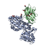
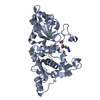
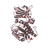
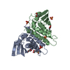
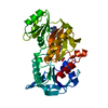
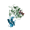
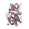
| Title | Crystal structure of a thermostable green fluorescent protein (TGP) with a synthetic nanobody (Sb44) | ||||||||||||
 Components Components |
| ||||||||||||
 Keywords Keywords | FLUORESCENT PROTEIN / complex / GFP / nanobody / single-chain antibody / sybody / synthetic antibody / TGP / thermostable green fluorescent protein | ||||||||||||
| Function / homology | ACETATE ION Function and homology information Function and homology information | ||||||||||||
| Biological species |  Galaxea fascicularis (invertebrata) Galaxea fascicularis (invertebrata) | ||||||||||||
| Method |  X-RAY DIFFRACTION / SYNCHROTRON / MOLECULAR REPLACEMENT / Resolution: 2.03 Å X-RAY DIFFRACTION / SYNCHROTRON / MOLECULAR REPLACEMENT / Resolution: 2.03 Å | ||||||||||||
 Authors Authors | Cai, H. / Yao, H. / Li, T. / Hutter, C. / Tang, Y. / Li, Y. / Seeger, M. / Li, D. | ||||||||||||
| Funding support |  China, 1items China, 1items
| ||||||||||||
 Citation Citation | Journal: Commun Biol / Year: 2020 Title: An improved fluorescent tag and its nanobodies for membrane protein expression, stability assay, and purification. Authors: Cai, H. / Yao, H. / Li, T. / Hutter, C.A.J. / Li, Y. / Tang, Y. / Seeger, M.A. / Li, D. | ||||||||||||
| History |
|
- Structure visualization
| Structure viewer | Molecule: MolmilJmol/JSmol |
|---|
- Downloads & links
-Download
| PDBx/mmCIF format | 6lz2.cif.gz | 199.9 KB | Display | PDBx/mmCIF format |
|---|---|---|---|---|
| PDB format | pdb6lz2.ent.gz | 126 KB | Display | PDB format |
| PDBx/mmJSON format | 6lz2.json.gz | Tree view | PDBx/mmJSON format | |
| Others |  Other downloads Other downloads |
-Validation report
| Arichive directory | https://data.pdbj.org/pub/pdb/validation_reports/lz/6lz2ftp://data.pdbj.org/pub/pdb/validation_reports/lz/6lz2 | HTTPS FTP |
|---|
-Related structure data


-Links
 PDBj
PDBj


- Assembly
| Deposited unit | 
| ||||||||||||
|---|---|---|---|---|---|---|---|---|---|---|---|---|---|
| 1 | 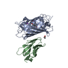
| ||||||||||||
| 2 | 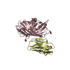
| ||||||||||||
| Unit cell |
|
-Components
-Protein / Antibody , 2 types, 4 molecules ACBD
| #1: Protein | Mass: 26699.957 Da / Num. of mol.: 2 Source method: isolated from a genetically manipulated source Details: uniprot A8CLT2 / Source: (gene. exp.) Galaxea fascicularis (invertebrata) / Gene: Green fluorescent GFP-like protein / Plasmid: pEC / Details (production host): pET based vector / Production host:  #2: Antibody | Mass: 15567.152 Da / Num. of mol.: 2 Source method: isolated from a genetically manipulated source Source: (gene. exp.) |
|---|
-Non-polymers , 5 types, 478 molecules 


| #3: Chemical |  Mass: 22.990 Da / Num. of mol.: 2 / Source method: obtained synthetically / Formula: Na Mass: 22.990 Da / Num. of mol.: 2 / Source method: obtained synthetically / Formula: Na#4: Chemical | ChemComp-ACT /  Mass: 59.044 Da / Num. of mol.: 4 / Source method: obtained synthetically / Formula: C2H3O2 Mass: 59.044 Da / Num. of mol.: 4 / Source method: obtained synthetically / Formula: C2H3O2#5: Chemical |  Mass: 92.094 Da / Num. of mol.: 2 / Source method: obtained synthetically / Formula: C3H8O3 Mass: 92.094 Da / Num. of mol.: 2 / Source method: obtained synthetically / Formula: C3H8O3#6: Chemical | ChemComp-TRS / |  Mass: 122.143 Da / Num. of mol.: 1 / Source method: obtained synthetically / Formula: C4H12NO3 / Comment: pH buffer*YM Mass: 122.143 Da / Num. of mol.: 1 / Source method: obtained synthetically / Formula: C4H12NO3 / Comment: pH buffer*YM#7: Water | ChemComp-HOH / | Mass: 18.015 Da / Num. of mol.: 469 / Source method: isolated from a natural source / Formula: H2O |
|---|
-Details
| Has ligand of interest | Y |
|---|---|
| Has protein modification | Y |
-Experimental details
-Experiment
| Experiment | Method: X-RAY DIFFRACTION / Number of used crystals: 1 |
|---|
- Sample preparation
| Crystal | Density Matthews: 2.35 Å3/Da / Density % sol: 47.7 % / Description: 0.2-mm plate clusters |
|---|---|
| Crystal grow | Temperature: 293 K / Method: vapor diffusion, sitting drop / pH: 6.5 Details: 0.2M ammonium acetate, 25 %(w/v) polyethylene glycol 3350, 0.1M Bis-Tris pH 6.5 |
-Data collection
| Diffraction | Mean temperature: 100 K / Serial crystal experiment: N |
|---|---|
| Diffraction source | Source: SYNCHROTRON / Site: SSRF / Beamline: BL19U1 / Wavelength: 0.9793 Å |
| Detector | Type: DECTRIS PILATUS3 S 6M / Detector: PIXEL / Date: Jan 3, 2020 |
| Radiation | Protocol: SINGLE WAVELENGTH / Monochromatic (M) / Laue (L): M / Scattering type: x-ray |
| Radiation wavelength | Wavelength: 0.9793 Å / Relative weight: 1 |
| Reflection | Resolution: 2.03→49.53 Å / Num. obs: 52046 / % possible obs: 99.3 % / Redundancy: 13.1 % / Biso Wilson estimate: 34.87 Å2 / CC1/2: 0.999 / Rmerge(I) obs: 0.127 / Rpim(I) all: 0.037 / Χ2: 0.94 / Net I/σ(I): 13.9 |
| Reflection shell | Resolution: 2.03→2.09 Å / Redundancy: 11.9 % / Rmerge(I) obs: 1.694 / Mean I/σ(I) obs: 1.6 / Num. unique obs: 3497 / CC1/2: 0.67 / CC star: 0.896 / Rpim(I) all: 0.504 / Χ2: 0.84 / % possible all: 91.3 |
- Processing
| Software |
| |||||||||||||||||||||||||||||||||||||||||||||||||||||||||||||||||||||||||||||||||||||||||||||||||||||||||
|---|---|---|---|---|---|---|---|---|---|---|---|---|---|---|---|---|---|---|---|---|---|---|---|---|---|---|---|---|---|---|---|---|---|---|---|---|---|---|---|---|---|---|---|---|---|---|---|---|---|---|---|---|---|---|---|---|---|---|---|---|---|---|---|---|---|---|---|---|---|---|---|---|---|---|---|---|---|---|---|---|---|---|---|---|---|---|---|---|---|---|---|---|---|---|---|---|---|---|---|---|---|---|---|---|---|---|
| Refinement | Method to determine structure: MOLECULAR REPLACEMENT Starting model: 4tza, 5m13 Resolution: 2.03→45.04 Å / SU ML: 0.2348 / Cross valid method: FREE R-VALUE / σ(F): 1.34 / Phase error: 23.3682
| |||||||||||||||||||||||||||||||||||||||||||||||||||||||||||||||||||||||||||||||||||||||||||||||||||||||||
| Solvent computation | Shrinkage radii: 0.9 Å / VDW probe radii: 1.11 Å | |||||||||||||||||||||||||||||||||||||||||||||||||||||||||||||||||||||||||||||||||||||||||||||||||||||||||
| Displacement parameters | Biso mean: 39.4 Å2 | |||||||||||||||||||||||||||||||||||||||||||||||||||||||||||||||||||||||||||||||||||||||||||||||||||||||||
| Refinement step | Cycle: LAST / Resolution: 2.03→45.04 Å
| |||||||||||||||||||||||||||||||||||||||||||||||||||||||||||||||||||||||||||||||||||||||||||||||||||||||||
| Refine LS restraints |
| |||||||||||||||||||||||||||||||||||||||||||||||||||||||||||||||||||||||||||||||||||||||||||||||||||||||||
| LS refinement shell |
|
