 Movie
Movie Controller

[English] 日本語
 Yorodumi
- PDB-1gsa: STRUCTURE OF GLUTATHIONE SYNTHETASE COMPLEXED WITH ADP AND GLUTATHIONE -
+ Open data

- Basic information
| Entry | Database: PDB / ID: 1gsa | ||||||
|---|---|---|---|---|---|---|---|
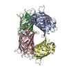
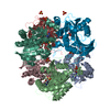
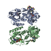
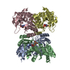
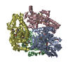
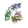
| Title | STRUCTURE OF GLUTATHIONE SYNTHETASE COMPLEXED WITH ADP AND GLUTATHIONE | ||||||
 Components Components | GLUTATHIONE SYNTHETASE | ||||||
 Keywords Keywords | LIGASE / GLUTATHIONE SYNTHETASE | ||||||
| Function / homology |  Function and homology information Function and homology informationglutathione synthase / glutathione synthase activity / glutathione biosynthetic process / protein homotetramerization / magnesium ion binding / ATP binding / identical protein binding / cytoplasm / cytosol Similarity search - Function | ||||||
| Biological species |  | ||||||
| Method |  X-RAY DIFFRACTION / SYNCHROTRON / Resolution: 2 Å X-RAY DIFFRACTION / SYNCHROTRON / Resolution: 2 Å | ||||||
 Authors Authors | Hara, T. / Kato, H. / Nishioka, T. / Katsube, Y. / Oda, J. | ||||||
 Citation Citation | Journal: Biochemistry / Year: 1996 Title: A pseudo-michaelis quaternary complex in the reverse reaction of a ligase: structure of Escherichia coli B glutathione synthetase complexed with ADP, glutathione, and sulfate at 2.0 A resolution. Authors: Hara, T. / Kato, H. / Katsube, Y. / Oda, J. #1: Journal: Biochemistry / Year: 1994Title: Flexible Loop that is Novel Catalytic Machinery in a Ligase. Atomic Structure and Function of the Loopless Glutathione Synthetase Authors: Kato, H. / Tanaka, T. / Yamaguchi, H. / Hara, T. / Nishioka, T. / Katsube, Y. / Oda, J. #2: Journal: J.Mol.Biol. / Year: 1993Title: Three-Dimensional Structure of the Glutathione Synthetase from Escherichia Coli B at 2.0 A Resolution Authors: Yamaguchi, H. / Kato, H. / Hata, Y. / Nishioka, T. / Kimura, A. / Oda, J. / Katsube, Y. #3: Journal: Agric.Biol.Chem. / Year: 1989Title: Overexpression of Glutathione Synthetase in Escherichia Coli Authors: Kato, H. / Kobayashi, M. / Murata, K. / Nishioka, T. / Oda, J. #4: Journal: J.Mol.Biol. / Year: 1989Title: Crystallization and Preliminary X-Ray Studies of Glutathione Synthetase from Escherichia Coli B Authors: Kato, H. / Yamaguchi, H. / Hata, Y. / Nishioka, T. / Katsube, Y. / Oda, J. #5: Journal: J.Biol.Chem. / Year: 1988Title: Role of Cysteine Residues in Glutathione Synthetase from Escherichia Coli B. Chemical Modification and Oligonucleotide Site-Directed Mutagenesis Authors: Kato, H. / Tanaka, T. / Nishioka, T. / Kimura, A. / Oda, J. | ||||||
| History |
|
- Structure visualization
| Structure viewer | Molecule: MolmilJmol/JSmol |
|---|
- Downloads & links
-Download
| PDBx/mmCIF format | 1gsa.cif.gz | 81.8 KB | Display | PDBx/mmCIF format |
|---|---|---|---|---|
| PDB format | pdb1gsa.ent.gz | 60.2 KB | Display | PDB format |
| PDBx/mmJSON format | 1gsa.json.gz | Tree view | PDBx/mmJSON format | |
| Others |  Other downloads Other downloads |
-Validation report
| Arichive directory | https://data.pdbj.org/pub/pdb/validation_reports/gs/1gsaftp://data.pdbj.org/pub/pdb/validation_reports/gs/1gsa | HTTPS FTP |
|---|
-Related structure data
| Similar structure data |
|---|


-Links
 PDBj
PDBj


- Assembly
| Deposited unit | 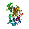
| ||||||||
|---|---|---|---|---|---|---|---|---|---|
| 1 | 
| ||||||||
| 2 | 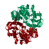
| ||||||||
| Unit cell |
|
-Components
-Protein , 1 types, 1 molecules A
| #1: Protein | Mass: 35601.824 Da / Num. of mol.: 1 Source method: isolated from a genetically manipulated source Source: (gene. exp.) |
|---|
-Non-polymers , 5 types, 140 molecules 


| #2: Chemical |  Mass: 24.305 Da / Num. of mol.: 2 / Source method: obtained synthetically / Formula: Mg Mass: 24.305 Da / Num. of mol.: 2 / Source method: obtained synthetically / Formula: Mg#3: Chemical | ChemComp-SO4 / |  Mass: 96.063 Da / Num. of mol.: 1 / Source method: obtained synthetically / Formula: SO4 Mass: 96.063 Da / Num. of mol.: 1 / Source method: obtained synthetically / Formula: SO4#4: Chemical | ChemComp-ADP / |  Mass: 427.201 Da / Num. of mol.: 1 / Source method: obtained synthetically / Formula: C10H15N5O10P2 / Comment: ADP, energy-carrying molecule*YM Mass: 427.201 Da / Num. of mol.: 1 / Source method: obtained synthetically / Formula: C10H15N5O10P2 / Comment: ADP, energy-carrying molecule*YM#5: Chemical | ChemComp-GSH / |  Mass: 307.323 Da / Num. of mol.: 1 / Source method: obtained synthetically / Formula: C10H17N3O6S Mass: 307.323 Da / Num. of mol.: 1 / Source method: obtained synthetically / Formula: C10H17N3O6S#6: Water | ChemComp-HOH / | Mass: 18.015 Da / Num. of mol.: 135 / Source method: isolated from a natural source / Formula: H2O |
|---|
-Experimental details
-Experiment
| Experiment | Method: X-RAY DIFFRACTION |
|---|
- Sample preparation
| Crystal | Density Matthews: 2.62 Å3/Da / Density % sol: 53 % | ||||||||||||||||||||||||||||||||||||
|---|---|---|---|---|---|---|---|---|---|---|---|---|---|---|---|---|---|---|---|---|---|---|---|---|---|---|---|---|---|---|---|---|---|---|---|---|---|
| Crystal grow | pH: 7.5 Details: CRYSTALLIZED IN TRIS-HCL BUFFER (PH 7.5). AMMONIUM SULFATE WAS USED AS PRECIPITANT. | ||||||||||||||||||||||||||||||||||||
| Crystal | *PLUS | ||||||||||||||||||||||||||||||||||||
| Crystal grow | *PLUS Temperature: 25 ℃ / Method: microdialysis | ||||||||||||||||||||||||||||||||||||
| Components of the solutions | *PLUS
|
-Data collection
| Diffraction source | Source: SYNCHROTRON / Site: Photon Factory  / Beamline: BL-6A / Wavelength: 1.04 / Beamline: BL-6A / Wavelength: 1.04 |
|---|---|
| Detector | Detector: FILM / Date: Nov 28, 1991 |
| Radiation | Monochromatic (M) / Laue (L): M / Scattering type: x-ray |
| Radiation wavelength | Wavelength: 1.04 Å / Relative weight: 1 |
| Reflection | Resolution: 1.8→45.1 Å / Num. obs: 23410 / % possible obs: 81.5 % / Observed criterion σ(I): 2 / Rmerge(I) obs: 0.058 |
| Reflection | *PLUS Num. measured all: 196511 |
- Processing
| Software |
| ||||||||||||||||||||||||||||||||||||||||||||||||||||||||||||
|---|---|---|---|---|---|---|---|---|---|---|---|---|---|---|---|---|---|---|---|---|---|---|---|---|---|---|---|---|---|---|---|---|---|---|---|---|---|---|---|---|---|---|---|---|---|---|---|---|---|---|---|---|---|---|---|---|---|---|---|---|---|
| Refinement | Resolution: 2→8 Å / σ(F): 2
| ||||||||||||||||||||||||||||||||||||||||||||||||||||||||||||
| Displacement parameters | Biso mean: 19.61 Å2 | ||||||||||||||||||||||||||||||||||||||||||||||||||||||||||||
| Refine analyze | Luzzati coordinate error obs: 0.22 Å | ||||||||||||||||||||||||||||||||||||||||||||||||||||||||||||
| Refinement step | Cycle: LAST / Resolution: 2→8 Å
| ||||||||||||||||||||||||||||||||||||||||||||||||||||||||||||
| Refine LS restraints |
| ||||||||||||||||||||||||||||||||||||||||||||||||||||||||||||
| Xplor file |
| ||||||||||||||||||||||||||||||||||||||||||||||||||||||||||||
| Software | *PLUS Name: X-PLOR / Version: 3 / Classification: refinement | ||||||||||||||||||||||||||||||||||||||||||||||||||||||||||||
| Refine LS restraints | *PLUS
|
