 Movie
Movie Controller

[English] 日本語
 Yorodumi
- PDB-2a8y: Crystal structure of 5'-deoxy-5'methylthioadenosine phosphorylase... -
+ Open data

- Basic information
| Entry | Database: PDB / ID: 2a8y | ||||||
|---|---|---|---|---|---|---|---|
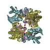
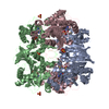
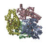
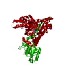
| Title | Crystal structure of 5'-deoxy-5'methylthioadenosine phosphorylase complexed with 5'-deoxy-5'methylthioadenosine and sulfate | ||||||
 Components Components | 5'-methylthioadenosine phosphorylase (mtaP) | ||||||
 Keywords Keywords | TRANSFERASE / alpha/beta / beta sheet / beta barrel | ||||||
| Function / homology |  Function and homology information Function and homology informationS-methyl-5'-thioadenosine phosphorylase / S-methyl-5-thioadenosine phosphorylase activity / : / purine ribonucleoside salvage / cytosol Similarity search - Function | ||||||
| Biological species |   Sulfolobus solfataricus (archaea) Sulfolobus solfataricus (archaea) | ||||||
| Method |  X-RAY DIFFRACTION / SYNCHROTRON / MOLECULAR REPLACEMENT / Resolution: 1.45 Å X-RAY DIFFRACTION / SYNCHROTRON / MOLECULAR REPLACEMENT / Resolution: 1.45 Å | ||||||
 Authors Authors | Zhang, Y. / Porcelli, M. / Cacciapuoti, G. / Ealick, S.E. | ||||||
 Citation Citation | Journal: J.Mol.Biol. / Year: 2006 Title: The crystal structure of 5'-deoxy-5'-methylthioadenosine phosphorylase II from Sulfolobus solfataricus, a thermophilic enzyme stabilized by intramolecular disulfide bonds. Authors: Zhang, Y. / Porcelli, M. / Cacciapuoti, G. / Ealick, S.E. | ||||||
| History |
|
- Structure visualization
| Structure viewer | Molecule: MolmilJmol/JSmol |
|---|
- Downloads & links
-Download
| PDBx/mmCIF format | 2a8y.cif.gz | 721.7 KB | Display | PDBx/mmCIF format |
|---|---|---|---|---|
| PDB format | pdb2a8y.ent.gz | 585.3 KB | Display | PDB format |
| PDBx/mmJSON format | 2a8y.json.gz | Tree view | PDBx/mmJSON format | |
| Others |  Other downloads Other downloads |
-Validation report
| Arichive directory | https://data.pdbj.org/pub/pdb/validation_reports/a8/2a8yftp://data.pdbj.org/pub/pdb/validation_reports/a8/2a8y | HTTPS FTP |
|---|
-Related structure data
| Related structure data |  1cg6S S: Starting model for refinement |
|---|---|
| Similar structure data |


-Links
 PDBj
PDBj- Assembly
| Deposited unit | 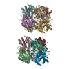
| ||||||||
|---|---|---|---|---|---|---|---|---|---|
| 1 | 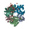
| ||||||||
| 2 | 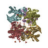
| ||||||||
| Unit cell |
|
-Components
| #1: Protein | Mass: 30177.795 Da / Num. of mol.: 12 Source method: isolated from a genetically manipulated source Source: (gene. exp.) Sulfolobus solfataricus (archaea) / Gene: SsMTAPII / Plasmid: pET-22b(+) / Species (production host): Escherichia coli / Production host:  References: UniProt: Q97W94, S-methyl-5'-thioadenosine phosphorylase #2: Chemical | ChemComp-SO4 /   Mass: 96.063 Da / Num. of mol.: 24 / Source method: obtained synthetically / Formula: SO4 Mass: 96.063 Da / Num. of mol.: 24 / Source method: obtained synthetically / Formula: SO4#3: Chemical | ChemComp-MTA /   Mass: 297.334 Da / Num. of mol.: 12 / Source method: obtained synthetically / Formula: C11H15N5O3S Mass: 297.334 Da / Num. of mol.: 12 / Source method: obtained synthetically / Formula: C11H15N5O3S#4: Water | ChemComp-HOH / |  Mass: 18.015 Da / Num. of mol.: 3405 / Source method: isolated from a natural source / Formula: H2O Mass: 18.015 Da / Num. of mol.: 3405 / Source method: isolated from a natural source / Formula: H2OHas protein modification | Y | |
|---|
-Experimental details
-Experiment
| Experiment | Method: X-RAY DIFFRACTION / Number of used crystals: 1 |
|---|
- Sample preparation
| Crystal | Density Matthews: 2.52 Å3/Da / Density % sol: 51.2 % |
|---|---|
| Crystal grow | Temperature: 295 K / Method: vapor diffusion, hanging drop / pH: 6.5 Details: ammonium sulfate, Bis-Tris, pH 6.5, VAPOR DIFFUSION, HANGING DROP, temperature 295K |
-Data collection
| Diffraction | Mean temperature: 100 K |
|---|---|
| Diffraction source | Source: SYNCHROTRON / Site: APS  / Beamline: 8-BM / Wavelength: 0.97946 Å / Beamline: 8-BM / Wavelength: 0.97946 Å |
| Detector | Type: ADSC QUANTUM 315 / Detector: CCD / Date: Feb 28, 2004 / Details: mirrors |
| Radiation | Monochromator: GRAPHITE / Protocol: SINGLE WAVELENGTH / Monochromatic (M) / Laue (L): M / Scattering type: x-ray |
| Radiation wavelength | Wavelength: 0.97946 Å / Relative weight: 1 |
| Reflection | Resolution: 1.45→500 Å / Num. all: 579262 / Num. obs: 579262 / % possible obs: 94.2 % / Observed criterion σ(F): 0 / Observed criterion σ(I): 0 / Redundancy: 1.6 % / Biso Wilson estimate: 14.3 Å2 / Rsym value: 0.06 / Net I/σ(I): 13.3 |
| Reflection shell | Resolution: 1.45→1.5 Å / Redundancy: 1 % / Mean I/σ(I) obs: 3.7 / Num. unique all: 55440 / Rsym value: 0.245 / % possible all: 90.1 |
- Processing
| Software |
| |||||||||||||||||||||||||
|---|---|---|---|---|---|---|---|---|---|---|---|---|---|---|---|---|---|---|---|---|---|---|---|---|---|---|
| Refinement | Method to determine structure: MOLECULAR REPLACEMENT Starting model: PDB entry 1CG6 Resolution: 1.45→48.27 Å / Rfactor Rfree error: 0.001 / Data cutoff high absF: 1148613.1 / Data cutoff low absF: 0 / Isotropic thermal model: RESTRAINED / Cross valid method: THROUGHOUT / σ(F): 0 / Stereochemistry target values: Engh & Huber
| |||||||||||||||||||||||||
| Solvent computation | Solvent model: FLAT MODEL / Bsol: 50.0119 Å2 / ksol: 0.362326 e/Å3 | |||||||||||||||||||||||||
| Displacement parameters | Biso mean: 18.4 Å2
| |||||||||||||||||||||||||
| Refine analyze |
| |||||||||||||||||||||||||
| Refinement step | Cycle: LAST / Resolution: 1.45→48.27 Å
| |||||||||||||||||||||||||
| Refine LS restraints |
| |||||||||||||||||||||||||
| LS refinement shell | Resolution: 1.45→1.54 Å / Rfactor Rfree error: 0.004 / Total num. of bins used: 6
| |||||||||||||||||||||||||
| Xplor file |
|
