 Movie
Movie Controller


+ Open data

- Basic information
| Entry | Database: PDB / ID: 3ja4 | ||||||
|---|---|---|---|---|---|---|---|
| Title | RNA-dependent RNA polymerases of transcribing cypoviruses | ||||||
 Components Components | RNA-dependent RNA polymerase | ||||||
 Keywords Keywords | TRANSFERASE / RNA-dependent RNA polymerases / transcribing cypoviruses / icosahedral virus | ||||||
| Function / homology |  Function and homology information Function and homology informationviral genome replication / RNA-directed RNA polymerase / RNA-directed RNA polymerase activity / RNA binding Similarity search - Function | ||||||
| Biological species |   Bombyx mori cypovirus 1 Bombyx mori cypovirus 1 | ||||||
| Method | ELECTRON MICROSCOPY / single particle reconstruction / cryo EM / Resolution: 4.8 Å | ||||||
 Authors Authors | Liu, H. / Cheng, L. | ||||||
 Citation Citation | Journal: Science / Year: 2015 Title: Cryo-EM shows the polymerase structures and a nonspooled genome within a dsRNA virus. Authors: Hongrong Liu / Lingpeng Cheng /  Abstract: Double-stranded RNA (dsRNA) viruses possess a segmented dsRNA genome and a number of RNA-dependent RNA polymerases (RdRps) enclosed in a capsid. Until now, the precise structures of genomes and RdRps ...Double-stranded RNA (dsRNA) viruses possess a segmented dsRNA genome and a number of RNA-dependent RNA polymerases (RdRps) enclosed in a capsid. Until now, the precise structures of genomes and RdRps within the capsids have been unknown. Here we report the structures of RdRps and associated RNAs within nontranscribing and transcribing cypoviruses (NCPV and TCPV, respectively), using a combination of cryo-electron microscopy (cryo-EM) and a symmetry-mismatch reconstruction method. The RdRps and associated RNAs appear to exhibit a pseudo-D3 symmetric organization in both NCPV and TCPV. However, the molecular interactions between RdRps and the genomic RNA were found to differ in these states. Our work provides insight into the mechanisms of the replication and transcription in dsRNA viruses and paves a way for structural determination of lower-symmetry complexes enclosed in higher-symmetry structures. | ||||||
| History |
|
- Structure visualization
| Movie |
Movie viewer |
|---|---|
| Structure viewer | Molecule: MolmilJmol/JSmol |
- Downloads & links
-Download
| PDBx/mmCIF format | 3ja4.cif.gz | 49.3 KB | Display | PDBx/mmCIF format |
|---|---|---|---|---|
| PDB format | pdb3ja4.ent.gz | 24.3 KB | Display | PDB format |
| PDBx/mmJSON format | 3ja4.json.gz | Tree view | PDBx/mmJSON format | |
| Others |  Other downloads Other downloads |
-Validation report
| Arichive directory | https://data.pdbj.org/pub/pdb/validation_reports/ja/3ja4ftp://data.pdbj.org/pub/pdb/validation_reports/ja/3ja4 | HTTPS FTP |
|---|
-Related structure data
| Related structure data | 6321MC 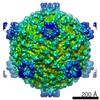 6322C  3ja5C M: map data used to model this data C: citing same article ( |
|---|---|
| Similar structure data |
-Links
 PDBj
PDBj- Assembly
| Deposited unit | 
|
|---|---|
| 1 | x 6
|
| 2 |
|
| 3 | 
|
| Symmetry | Point symmetry: (Schoenflies symbol: D3 (2x3 fold dihedral)) |
-Components
| #1: Protein | Mass: 138830.109 Da / Num. of mol.: 1 / Source method: isolated from a natural source / Source: (natural) Bombyx mori cypovirus 1 / References: UniProt: Q6RJR5 |
|---|
-Experimental details
-Experiment
| Experiment | Method: ELECTRON MICROSCOPY |
|---|---|
| EM experiment | Aggregation state: PARTICLE / 3D reconstruction method: single particle reconstruction |
- Sample preparation
| Component | Name: transcribing cypoviruses / Type: VIRUS |
|---|---|
| Specimen | Embedding applied: NO / Shadowing applied: NO / Staining applied: NO / Vitrification applied: YES |
| Vitrification | Instrument: FEI VITROBOT MARK IV / Cryogen name: ETHANE |
- Electron microscopy imaging
| Experimental equipment |  Model: Titan Krios / Image courtesy: FEI Company |
|---|---|
| Microscopy | Model: FEI TITAN KRIOS / Date: Oct 10, 2012 |
| Electron gun | Electron source:  FIELD EMISSION GUN / Accelerating voltage: 300 kV / Illumination mode: OTHER FIELD EMISSION GUN / Accelerating voltage: 300 kV / Illumination mode: OTHER |
| Electron lens | Mode: BRIGHT FIELD / Nominal magnification: 75000 X / Nominal defocus max: 3 nm / Nominal defocus min: 1 nm |
| Image recording | Film or detector model: GATAN ULTRASCAN 4000 (4k x 4k) |
- Processing
| Symmetry | Point symmetry: C1 (asymmetric) | ||||||||||||
|---|---|---|---|---|---|---|---|---|---|---|---|---|---|
| 3D reconstruction | Method: Symmetry-mismatch reconstruction of icosahedral virus Resolution: 4.8 Å / Num. of particles: 36000 / Symmetry type: POINT | ||||||||||||
| Refinement step | Cycle: LAST
|
