 Movie
Movie Controller


+ Open data

- Basic information
| Entry | Database: PDB / ID: 1kfu | |||||||||
|---|---|---|---|---|---|---|---|---|---|---|
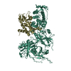
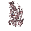
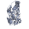
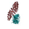
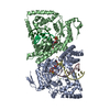
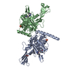
| Title | Crystal Structure of Human m-Calpain Form II | |||||||||
 Components Components |
| |||||||||
 Keywords Keywords | HYDROLASE / REGULATION / PAPAIN-LIKE / THIOL-PROTEASE | |||||||||
| Function / homology |  Function and homology information Function and homology informationcalpain-2 / calpain complex / positive regulation of phosphatidylcholine biosynthetic process / protein catabolic process at postsynapse / calcium-dependent cysteine-type endopeptidase activity / perinuclear endoplasmic reticulum / Formation of the cornified envelope / regulation of interleukin-6 production / positive regulation of myoblast fusion / cortical actin cytoskeleton ...calpain-2 / calpain complex / positive regulation of phosphatidylcholine biosynthetic process / protein catabolic process at postsynapse / calcium-dependent cysteine-type endopeptidase activity / perinuclear endoplasmic reticulum / Formation of the cornified envelope / regulation of interleukin-6 production / positive regulation of myoblast fusion / cortical actin cytoskeleton / Deregulated CDK5 triggers multiple neurodegenerative pathways in Alzheimer's disease models / regulation of cytoskeleton organization / vascular endothelial cell response to oscillatory fluid shear stress / protein autoprocessing / pseudopodium / behavioral response to pain / regulation of macroautophagy / response to mechanical stimulus / synaptic vesicle endocytosis / vascular endothelial cell response to laminar fluid shear stress / cellular response to interferon-beta / Degradation of the extracellular matrix / cytoskeletal protein binding / cysteine-type peptidase activity / positive regulation of cardiac muscle cell apoptotic process / Turbulent (oscillatory, disturbed) flow shear stress activates signaling by PIEZO1 and integrins in endothelial cells / : / cellular response to amino acid stimulus / response to hydrogen peroxide / female pregnancy / cellular response to lipopolysaccharide / presynapse / High laminar flow shear stress activates signaling by PIEZO1 and PECAM1:CDH5:KDR in endothelial cells / response to hypoxia / lysosome / postsynapse / membrane raft / external side of plasma membrane / focal adhesion / neuronal cell body / calcium ion binding / positive regulation of cell population proliferation / dendrite / chromatin / protein-containing complex binding / Golgi apparatus / enzyme binding / endoplasmic reticulum / proteolysis / extracellular exosome / membrane / nucleus / plasma membrane / cytosol / cytoplasm Similarity search - Function | |||||||||
| Biological species |  Homo sapiens (human) Homo sapiens (human) | |||||||||
| Method |  X-RAY DIFFRACTION / SYNCHROTRON / MAD / Resolution: 2.5 Å X-RAY DIFFRACTION / SYNCHROTRON / MAD / Resolution: 2.5 Å | |||||||||
 Authors Authors | Strobl, S. / Fernandez-Catalan, C. / Braun, M. / Huber, R. / Masumoto, H. / Nakagawa, K. / Irie, A. / Sorimachi, H. / Bourenkow, G. / Bartunik, H. ...Strobl, S. / Fernandez-Catalan, C. / Braun, M. / Huber, R. / Masumoto, H. / Nakagawa, K. / Irie, A. / Sorimachi, H. / Bourenkow, G. / Bartunik, H. / Suzuki, K. / Bode, W. | |||||||||
 Citation Citation | Journal: Proc.Natl.Acad.Sci.USA / Year: 2000 Title: The crystal structure of calcium-free human m-calpain suggests an electrostatic switch mechanism for activation by calcium. Authors: Strobl, S. / Fernandez-Catalan, C. / Braun, M. / Huber, R. / Masumoto, H. / Nakagawa, K. / Irie, A. / Sorimachi, H. / Bourenkow, G. / Bartunik, H. / Suzuki, K. / Bode, W. #1: Journal: Acta Crystallogr.,Sect.D / Year: 2000Title: Crystallization and preliminary X-ray analysis of recombinant full-length m-calpain Authors: Masumoto, H. / Nakagawa, K. / Irie, S. / Sorimachi, H. / Suzuki, K. / Bourenkow, G. / Bartunik, H. / Fernandez-Catalan, C. / Bode, W. / Strobl, S. #2: Journal: Biol.Chem. / Year: 2001Title: Structural basis for possible calcium-induced activation mechanisms of calpains Authors: Reverter, D. / Strobl, S. / Fernandez-Catalan, C. / Sorimachi, H. / Suzuki, K. / Bode, W. #3: Journal: Trends Cardiovasc.Med. / Year: 2001Title: The structure of calcium-free human m-calpain Authors: Reverter, D. / Sorimachi, H. / Bode, W. | |||||||||
| History |
|
- Structure visualization
| Structure viewer | Molecule: MolmilJmol/JSmol |
|---|
- Downloads & links
-Download
| PDBx/mmCIF format | 1kfu.cif.gz | 195.5 KB | Display | PDBx/mmCIF format |
|---|---|---|---|---|
| PDB format | pdb1kfu.ent.gz | 152.2 KB | Display | PDB format |
| PDBx/mmJSON format | 1kfu.json.gz | Tree view | PDBx/mmJSON format | |
| Others |  Other downloads Other downloads |
-Validation report
| Arichive directory | https://data.pdbj.org/pub/pdb/validation_reports/kf/1kfuftp://data.pdbj.org/pub/pdb/validation_reports/kf/1kfu | HTTPS FTP |
|---|
-Related structure data


-Links
 PDBj
PDBj


- Assembly
| Deposited unit | 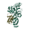
| ||||||||||
|---|---|---|---|---|---|---|---|---|---|---|---|
| 1 |
| ||||||||||
| Unit cell |
|
-Components
| #1: Protein | Mass: 79968.023 Da / Num. of mol.: 1 / Fragment: CATALYTIC SUBUNIT Source method: isolated from a genetically manipulated source Source: (gene. exp.) Homo sapiens (human) / Production host:   Spodoptera frugiperda (fall armyworm) / References: UniProt: P17655, calpain-2 Spodoptera frugiperda (fall armyworm) / References: UniProt: P17655, calpain-2 |
|---|---|
| #2: Protein | Mass: 21263.859 Da / Num. of mol.: 1 / Fragment: REGULATORY SUBUNIT Source method: isolated from a genetically manipulated source Source: (gene. exp.) Homo sapiens (human) / Production host: Spodoptera frugiperda (fall armyworm) / References: UniProt: P04632 |
| #3: Water | ChemComp-HOH /  Mass: 18.015 Da / Num. of mol.: 411 / Source method: isolated from a natural source / Formula: H2O Mass: 18.015 Da / Num. of mol.: 411 / Source method: isolated from a natural source / Formula: H2O |
-Experimental details
-Experiment
| Experiment | Method: X-RAY DIFFRACTION / Number of used crystals: 1 |
|---|
- Sample preparation
| Crystal | Density Matthews: 2.79 Å3/Da / Density % sol: 55.95 % | ||||||||||||||||||||||||||||||||||||||||||||||||||||||||||||||||||||||
|---|---|---|---|---|---|---|---|---|---|---|---|---|---|---|---|---|---|---|---|---|---|---|---|---|---|---|---|---|---|---|---|---|---|---|---|---|---|---|---|---|---|---|---|---|---|---|---|---|---|---|---|---|---|---|---|---|---|---|---|---|---|---|---|---|---|---|---|---|---|---|---|
| Crystal grow | Temperature: 293 K / Method: vapor diffusion, sitting drop / pH: 7.5 Details: PEG 10000, isopropanol, guanidinium chloride, pH 7.5, VAPOR DIFFUSION, SITTING DROP at 293K, VAPOR DIFFUSION, SITTING DROP | ||||||||||||||||||||||||||||||||||||||||||||||||||||||||||||||||||||||
| Crystal grow | *PLUS Method: vapor diffusionDetails: Masumoto, H., (2000) Acta Crystallogr., Sect.D, 56, 73. | ||||||||||||||||||||||||||||||||||||||||||||||||||||||||||||||||||||||
| Components of the solutions | *PLUS
|
-Data collection
| Diffraction | Mean temperature: 100 K |
|---|---|
| Diffraction source | Source: SYNCHROTRON / Site: EMBL/DESY, HAMBURG  / Beamline: BW7B / Beamline: BW7B |
| Detector | Type: MARRESEARCH / Detector: CCD / Date: Oct 11, 1998 |
| Radiation | Protocol: MAD / Monochromatic (M) / Laue (L): M / Scattering type: x-ray |
| Radiation wavelength | Relative weight: 1 |
| Reflection | Highest resolution: 2.3 Å / Num. all: 47236 / Num. obs: 47236 / % possible obs: 94.8 % / Rmerge(I) obs: 0.045 |
| Reflection | *PLUS Num. measured all: 400680 |
- Processing
| Software |
| ||||||||||||||||
|---|---|---|---|---|---|---|---|---|---|---|---|---|---|---|---|---|---|
| Refinement | Method to determine structure: MAD / Resolution: 2.5→30 Å / σ(F): 2 / Stereochemistry target values: Engh & Huber
| ||||||||||||||||
| Displacement parameters | Biso mean: 19 Å2 | ||||||||||||||||
| Refinement step | Cycle: LAST / Resolution: 2.5→30 Å
| ||||||||||||||||
| Refine LS restraints |
| ||||||||||||||||
| Software | *PLUS Name: CNS / Classification: refinement | ||||||||||||||||
| Refinement | *PLUS Lowest resolution: 30 Å / Num. reflection obs: 38544 / σ(F): 2 / % reflection Rfree: 4 % / Rfactor obs: 0.206 / Rfactor Rfree: 0.266 | ||||||||||||||||
| Solvent computation | *PLUS | ||||||||||||||||
| Displacement parameters | *PLUS Biso mean: 19 Å2 | ||||||||||||||||
| Refine LS restraints | *PLUS
|
