 Movie
Movie Controller

[English] 日本語
 Yorodumi
- PDB-7btk: E.coli beta-galactosidase (E537Q) in complex with fluorescent pro... -
+ Open data

- Basic information
| Entry | Database: PDB / ID: 7btk | |||||||||||||||
|---|---|---|---|---|---|---|---|---|---|---|---|---|---|---|---|---|
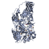
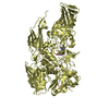
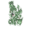
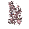
| Title | E.coli beta-galactosidase (E537Q) in complex with fluorescent probe KSA01 | |||||||||||||||
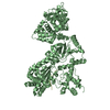
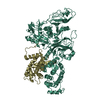
 Components Components | Beta-galactosidase | |||||||||||||||
 Keywords Keywords | HYDROLASE / beta_galactosidase / fluorescent probe | |||||||||||||||
| Function / homology |  Function and homology information Function and homology informationalkali metal ion binding / lactose catabolic process / beta-galactosidase complex / beta-galactosidase / beta-galactosidase activity / carbohydrate binding / magnesium ion binding / identical protein binding Similarity search - Function | |||||||||||||||
| Biological species |  | |||||||||||||||
| Method |  X-RAY DIFFRACTION / SYNCHROTRON / MOLECULAR REPLACEMENT / Resolution: 2.7 Å X-RAY DIFFRACTION / SYNCHROTRON / MOLECULAR REPLACEMENT / Resolution: 2.7 Å | |||||||||||||||
 Authors Authors | Chen, X. / Hu, Y.L. / Liu, Q.M. / Gao, Y. / Yuan, R. / Guo, Y. | |||||||||||||||
| Funding support |  China, 4items China, 4items
| |||||||||||||||
 Citation Citation | Journal: Angew.Chem.Int.Ed.Engl. / Year: 2021 Title: Two-Dimensional Design Strategy to Construct Smart Fluorescent Probes for the Precise Tracking of Senescence. Authors: Gao, Y. / Hu, Y. / Liu, Q. / Li, X. / Li, X. / Kim, C.Y. / James, T.D. / Li, J. / Chen, X. / Guo, Y. | |||||||||||||||
| History |
|
- Structure visualization
| Structure viewer | Molecule: MolmilJmol/JSmol |
|---|
- Downloads & links
-Download
| PDBx/mmCIF format | 7btk.cif.gz | 847.9 KB | Display | PDBx/mmCIF format |
|---|---|---|---|---|
| PDB format | pdb7btk.ent.gz | 680.7 KB | Display | PDB format |
| PDBx/mmJSON format | 7btk.json.gz | Tree view | PDBx/mmJSON format | |
| Others |  Other downloads Other downloads |
-Validation report
| Arichive directory | https://data.pdbj.org/pub/pdb/validation_reports/bt/7btkftp://data.pdbj.org/pub/pdb/validation_reports/bt/7btk | HTTPS FTP |
|---|
-Related structure data
| Related structure data |  7brsC  6kuzS S: Starting model for refinement C: citing same article ( |
|---|---|
| Similar structure data |
-Links
 PDBj
PDBj


- Assembly
| Deposited unit | 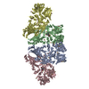
| ||||||||||||||||||||||||||||||||||||||||||||||||||||||||||||||||||||||||||||||||||||||||||||||||||||||||||||||||||||||||||||||||||||||||||||||||||||||
|---|---|---|---|---|---|---|---|---|---|---|---|---|---|---|---|---|---|---|---|---|---|---|---|---|---|---|---|---|---|---|---|---|---|---|---|---|---|---|---|---|---|---|---|---|---|---|---|---|---|---|---|---|---|---|---|---|---|---|---|---|---|---|---|---|---|---|---|---|---|---|---|---|---|---|---|---|---|---|---|---|---|---|---|---|---|---|---|---|---|---|---|---|---|---|---|---|---|---|---|---|---|---|---|---|---|---|---|---|---|---|---|---|---|---|---|---|---|---|---|---|---|---|---|---|---|---|---|---|---|---|---|---|---|---|---|---|---|---|---|---|---|---|---|---|---|---|---|---|---|---|---|
| 1 | 
| ||||||||||||||||||||||||||||||||||||||||||||||||||||||||||||||||||||||||||||||||||||||||||||||||||||||||||||||||||||||||||||||||||||||||||||||||||||||
| 2 | 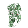
| ||||||||||||||||||||||||||||||||||||||||||||||||||||||||||||||||||||||||||||||||||||||||||||||||||||||||||||||||||||||||||||||||||||||||||||||||||||||
| Unit cell |
| ||||||||||||||||||||||||||||||||||||||||||||||||||||||||||||||||||||||||||||||||||||||||||||||||||||||||||||||||||||||||||||||||||||||||||||||||||||||
| Noncrystallographic symmetry (NCS) | NCS domain:
NCS domain segments: Component-ID: _ / Beg auth comp-ID: ILE / Beg label comp-ID: ILE / Refine code: _
NCS ensembles :
|
-Components
-Protein , 1 types, 4 molecules ABCD
| #1: Protein | Mass: 116688.547 Da / Num. of mol.: 4 / Mutation: E537Q Source method: isolated from a genetically manipulated source Source: (gene. exp.) |
|---|
-Non-polymers , 6 types, 1181 molecules 


| #2: Chemical | ChemComp-F6L /  Mass: 560.614 Da / Num. of mol.: 4 / Source method: obtained synthetically / Formula: C32H34NO8 / Feature type: SUBJECT OF INVESTIGATION Mass: 560.614 Da / Num. of mol.: 4 / Source method: obtained synthetically / Formula: C32H34NO8 / Feature type: SUBJECT OF INVESTIGATION#3: Chemical | ChemComp-MG /  Mass: 24.305 Da / Num. of mol.: 7 / Source method: obtained synthetically / Formula: Mg Mass: 24.305 Da / Num. of mol.: 7 / Source method: obtained synthetically / Formula: Mg#4: Chemical | ChemComp-NA /  Mass: 22.990 Da / Num. of mol.: 7 / Source method: obtained synthetically / Formula: Na Mass: 22.990 Da / Num. of mol.: 7 / Source method: obtained synthetically / Formula: Na#5: Chemical | ChemComp-DMS /  Mass: 78.133 Da / Num. of mol.: 38 / Source method: obtained synthetically / Formula: C2H6OS / Comment: DMSO, precipitant*YM Mass: 78.133 Da / Num. of mol.: 38 / Source method: obtained synthetically / Formula: C2H6OS / Comment: DMSO, precipitant*YM#6: Chemical | ChemComp-GOL / |  Mass: 92.094 Da / Num. of mol.: 1 / Source method: obtained synthetically / Formula: C3H8O3 Mass: 92.094 Da / Num. of mol.: 1 / Source method: obtained synthetically / Formula: C3H8O3#7: Water | ChemComp-HOH / | Mass: 18.015 Da / Num. of mol.: 1124 / Source method: isolated from a natural source / Formula: H2O |
|---|
-Details
| Has ligand of interest | Y |
|---|---|
| Has protein modification | N |
-Experimental details
-Experiment
| Experiment | Method: X-RAY DIFFRACTION / Number of used crystals: 1 |
|---|
- Sample preparation
| Crystal | Density Matthews: 2.78 Å3/Da / Density % sol: 55.71 % |
|---|---|
| Crystal grow | Temperature: 291 K / Method: vapor diffusion Details: 100 mM Bis-Tris, pH = 5.9, 200 mM NaCl, 200 mM MgCl2, 10% PEG8000, 10% glycerol, 0.1 M guanidine hydrochloride and 10 mM DTT |
-Data collection
| Diffraction | Mean temperature: 100 K / Serial crystal experiment: N |
|---|---|
| Diffraction source | Source: SYNCHROTRON / Site: SSRF / Beamline: BL18U1 / Wavelength: 0.9793 Å |
| Detector | Type: DECTRIS PILATUS3 S 6M / Detector: PIXEL / Date: Jan 13, 2020 |
| Radiation | Protocol: SINGLE WAVELENGTH / Monochromatic (M) / Laue (L): M / Scattering type: x-ray |
| Radiation wavelength | Wavelength: 0.9793 Å / Relative weight: 1 |
| Reflection | Resolution: 2.7→19.87 Å / Num. obs: 140397 / % possible obs: 99.6 % / Redundancy: 6.8 % / CC1/2: 0.991 / Rmerge(I) obs: 0.164 / Rpim(I) all: 0.068 / Rrim(I) all: 0.178 / Net I/σ(I): 9.5 |
| Reflection shell | Resolution: 2.7→2.75 Å / Redundancy: 7.1 % / Rmerge(I) obs: 0.737 / Mean I/σ(I) obs: 3.4 / Num. unique obs: 6952 / CC1/2: 0.838 / Rpim(I) all: 0.423 / Rrim(I) all: 0.797 / % possible all: 99.5 |
- Processing
| Software |
| |||||||||||||||||||||||||||||||||||||||||||||||||||||||||||||||||
|---|---|---|---|---|---|---|---|---|---|---|---|---|---|---|---|---|---|---|---|---|---|---|---|---|---|---|---|---|---|---|---|---|---|---|---|---|---|---|---|---|---|---|---|---|---|---|---|---|---|---|---|---|---|---|---|---|---|---|---|---|---|---|---|---|---|---|
| Refinement | Method to determine structure: MOLECULAR REPLACEMENT Starting model: 6KUZ Resolution: 2.7→19.87 Å / Cor.coef. Fo:Fc: 0.913 / Cor.coef. Fo:Fc free: 0.903 / SU B: 11.958 / SU ML: 0.243 / Cross valid method: THROUGHOUT / σ(F): 0 / ESU R Free: 0.324 / Stereochemistry target values: MAXIMUM LIKELIHOOD Details: HYDROGENS HAVE BEEN ADDED IN THE RIDING POSITIONS U VALUES : REFINED INDIVIDUALLY
| |||||||||||||||||||||||||||||||||||||||||||||||||||||||||||||||||
| Solvent computation | Ion probe radii: 0.8 Å / Shrinkage radii: 0.8 Å / VDW probe radii: 1.2 Å / Solvent model: MASK | |||||||||||||||||||||||||||||||||||||||||||||||||||||||||||||||||
| Displacement parameters | Biso max: 162.07 Å2 / Biso mean: 35.665 Å2 / Biso min: 0.5 Å2
| |||||||||||||||||||||||||||||||||||||||||||||||||||||||||||||||||
| Refinement step | Cycle: final / Resolution: 2.7→19.87 Å
| |||||||||||||||||||||||||||||||||||||||||||||||||||||||||||||||||
| Refine LS restraints |
| |||||||||||||||||||||||||||||||||||||||||||||||||||||||||||||||||
| Refine LS restraints NCS | Refine-ID: X-RAY DIFFRACTION / Type: interatomic distance / Weight position: 0.05
| |||||||||||||||||||||||||||||||||||||||||||||||||||||||||||||||||
| LS refinement shell | Resolution: 2.7→2.77 Å / Rfactor Rfree error: 0 / Total num. of bins used: 20
|
