 Movie
Movie Controller

[English] 日本語
 Yorodumi
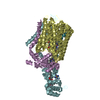
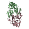
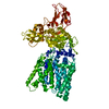
- PDB-6bvg: Crystal structure of bcMalT T280C-E54C crosslinked by divalent mercury -
+ Open data

- Basic information
| Entry | Database: PDB / ID: 6bvg | |||||||||
|---|---|---|---|---|---|---|---|---|---|---|
| Title | Crystal structure of bcMalT T280C-E54C crosslinked by divalent mercury | |||||||||
 Components Components | Protein-N(Pi)-phosphohistidine-sugar phosphotransferase (Enzyme II of the phosphotransferase system) (PTS system glucose-specific IIBC component) | |||||||||
 Keywords Keywords | TRANSPORT PROTEIN / EIIC maltose transporter | |||||||||
| Function / homology |  Function and homology information Function and homology informationprotein-Npi-phosphohistidine-sugar phosphotransferase / protein-phosphocysteine-sugar phosphotransferase activity / protein-N(PI)-phosphohistidine-carbohydrate phosphotransferase activity / phosphoenolpyruvate-dependent sugar phosphotransferase system / kinase activity / plasma membrane Similarity search - Function | |||||||||
| Biological species |  | |||||||||
| Method |  X-RAY DIFFRACTION / SYNCHROTRON / MOLECULAR REPLACEMENT / Resolution: 3.2 Å X-RAY DIFFRACTION / SYNCHROTRON / MOLECULAR REPLACEMENT / Resolution: 3.2 Å | |||||||||
 Authors Authors | Ren, Z. / Zhou, M. | |||||||||
| Funding support |  United States, 1items United States, 1items
| |||||||||
 Citation Citation | Journal: Proc. Natl. Acad. Sci. U.S.A. / Year: 2018 Title: Structure of an EIIC sugar transporter trapped in an inward-facing conformation. Authors: Ren, Z. / Lee, J. / Moosa, M.M. / Nian, Y. / Hu, L. / Xu, Z. / McCoy, J.G. / Ferreon, A.C.M. / Im, W. / Zhou, M. | |||||||||
| History |
|
- Structure visualization
| Structure viewer | Molecule: MolmilJmol/JSmol |
|---|
- Downloads & links
-Download
| PDBx/mmCIF format | 6bvg.cif.gz | 180.6 KB | Display | PDBx/mmCIF format |
|---|---|---|---|---|
| PDB format | pdb6bvg.ent.gz | 143.2 KB | Display | PDB format |
| PDBx/mmJSON format | 6bvg.json.gz | Tree view | PDBx/mmJSON format | |
| Others |  Other downloads Other downloads |
-Validation report
| Arichive directory | https://data.pdbj.org/pub/pdb/validation_reports/bv/6bvgftp://data.pdbj.org/pub/pdb/validation_reports/bv/6bvg | HTTPS FTP |
|---|
-Related structure data
| Related structure data |  5iwsS S: Starting model for refinement |
|---|---|
| Similar structure data |


-Links
 PDBj
PDBj


- Assembly
| Deposited unit | 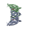
| ||||||||
|---|---|---|---|---|---|---|---|---|---|
| 1 |
| ||||||||
| Unit cell |
|
-Components
| #1: Protein | Mass: 48820.465 Da / Num. of mol.: 2 / Fragment: residues 3-451 / Mutation: T280C, E54C Source method: isolated from a genetically manipulated source Source: (gene. exp.) Strain: ZK / E33L / Gene: ptsG, BCE33L0344 / Plasmid: pMCSG28 / Production host: References: UniProt: Q63GK8, protein-Npi-phosphohistidine-sugar phosphotransferase #2: Polysaccharide |   Source method: isolated from a genetically manipulated source Details: oligosaccharide / References: alpha-maltose #3: Chemical |   Mass: 200.590 Da / Num. of mol.: 2 / Source method: obtained synthetically / Formula: Hg Mass: 200.590 Da / Num. of mol.: 2 / Source method: obtained synthetically / Formula: Hg |
|---|
-Experimental details
-Experiment
| Experiment | Method: X-RAY DIFFRACTION / Number of used crystals: 1 |
|---|
- Sample preparation
| Crystal | Density Matthews: 3.71 Å3/Da / Density % sol: 66.84 % |
|---|---|
| Crystal grow | Temperature: 277 K / Method: vapor diffusion, sitting drop Details: 100 mM Glycine, pH 8.8-9.1, 33-38% PEG 400, 50 mM CoCl2 and 10-20 mM spermine tetrahydrochloride PH range: 8.8-9.2 |
-Data collection
| Diffraction | Mean temperature: 100 K |
|---|---|
| Diffraction source | Source: SYNCHROTRON / Site: APS / Beamline: 17-ID / Wavelength: 0.9787 Å |
| Detector | Type: DECTRIS PILATUS 6M-F / Detector: PIXEL / Date: Feb 18, 2017 |
| Radiation | Monochromator: Si(111) / Protocol: SINGLE WAVELENGTH / Monochromatic (M) / Laue (L): M / Scattering type: x-ray |
| Radiation wavelength | Wavelength: 0.9787 Å / Relative weight: 1 |
| Reflection | Resolution: 3.2→29.42 Å / Num. obs: 24635 / % possible obs: 96.76 % / Redundancy: 4.5 % / Biso Wilson estimate: 37.47 Å2 / Rmerge(I) obs: 0.115 / Rpim(I) all: 0.058 / Rrim(I) all: 0.13 / Χ2: 0.998 / Net I/σ(I): 12.16 |
| Reflection shell | Resolution: 3.2→3.31 Å / Redundancy: 4.4 % / Rmerge(I) obs: 0.75 / Mean I/σ(I) obs: 1.8 / Num. unique obs: 2330 / CC1/2: 0.817 / Rpim(I) all: 0.384 / Rrim(I) all: 0.85 / Χ2: 0.884 / % possible all: 93.5 |
- Processing
| Software |
| ||||||||||||||||||||||||||||||||||||||||||||||||||||||||||||||||||||||
|---|---|---|---|---|---|---|---|---|---|---|---|---|---|---|---|---|---|---|---|---|---|---|---|---|---|---|---|---|---|---|---|---|---|---|---|---|---|---|---|---|---|---|---|---|---|---|---|---|---|---|---|---|---|---|---|---|---|---|---|---|---|---|---|---|---|---|---|---|---|---|---|
| Refinement | Method to determine structure: MOLECULAR REPLACEMENT Starting model: 5IWS Resolution: 3.2→29.42 Å / SU ML: 0.4 / Cross valid method: FREE R-VALUE / σ(F): 1.34 / Phase error: 38.28 / Stereochemistry target values: ML
| ||||||||||||||||||||||||||||||||||||||||||||||||||||||||||||||||||||||
| Solvent computation | Shrinkage radii: 0.9 Å / VDW probe radii: 1.11 Å / Solvent model: FLAT BULK SOLVENT MODEL | ||||||||||||||||||||||||||||||||||||||||||||||||||||||||||||||||||||||
| Refinement step | Cycle: LAST / Resolution: 3.2→29.42 Å
| ||||||||||||||||||||||||||||||||||||||||||||||||||||||||||||||||||||||
| Refine LS restraints |
| ||||||||||||||||||||||||||||||||||||||||||||||||||||||||||||||||||||||
| LS refinement shell |
|
