 Movie
Movie Controller

[English] 日本語
 Yorodumi
- PDB-1pww: Crystal structure of Anthrax Lethal Factor active site mutant pro... -
+ Open data

- Basic information
| Entry | Database: PDB / ID: 1pww | ||||||
|---|---|---|---|---|---|---|---|
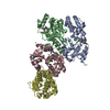
| Title | Crystal structure of Anthrax Lethal Factor active site mutant protein complexed with an optimised peptide substrate in the presence of zinc. | ||||||
 Components Components |
| ||||||
 Keywords Keywords | HYDROLASE / Anthrax Toxin / Lethal Factor (active site mutant) / optimised peptide substrate | ||||||
| Function / homology |  Function and homology information Function and homology informationanthrax lethal factor endopeptidase / host cell cytosol / Uptake and function of anthrax toxins / metalloendopeptidase activity / metallopeptidase activity / toxin activity / proteolysis / extracellular region / zinc ion binding Similarity search - Function | ||||||
| Biological species |  | ||||||
| Method |  X-RAY DIFFRACTION / SYNCHROTRON / MOLECULAR REPLACEMENT / Resolution: 2.8 Å X-RAY DIFFRACTION / SYNCHROTRON / MOLECULAR REPLACEMENT / Resolution: 2.8 Å | ||||||
 Authors Authors | Wong, T.Y. / Schwarzenbacher, R. / Liddington, R.C. | ||||||
 Citation Citation | Journal: Nat.Struct.Mol.Biol. / Year: 2004 Title: The structural basis for substrate and inhibitor selectivity of the anthrax lethal factor. Authors: Turk, B.E. / Wong, T.Y. / Schwarzenbacher, R. / Jarrell, E.T. / Leppla, S.H. / Collier, R.J. / Liddington, R.C. / Cantley, L.C. | ||||||
| History |
|
- Structure visualization
| Structure viewer | Molecule: MolmilJmol/JSmol |
|---|
- Downloads & links
-Download
| PDBx/mmCIF format | 1pww.cif.gz | 306.3 KB | Display | PDBx/mmCIF format |
|---|---|---|---|---|
| PDB format | pdb1pww.ent.gz | 248.1 KB | Display | PDB format |
| PDBx/mmJSON format | 1pww.json.gz | Tree view | PDBx/mmJSON format | |
| Others |  Other downloads Other downloads |
-Validation report
| Arichive directory | https://data.pdbj.org/pub/pdb/validation_reports/pw/1pwwftp://data.pdbj.org/pub/pdb/validation_reports/pw/1pww | HTTPS FTP |
|---|
-Related structure data
| Related structure data |  1pwqC  1pwuC  1pwvC  1j7nS C: citing same article ( S: Starting model for refinement |
|---|---|
| Similar structure data |


-Links
 PDBj
PDBj


- Assembly
| Deposited unit | 
| ||||||||
|---|---|---|---|---|---|---|---|---|---|
| 1 | 
| ||||||||
| 2 | 
| ||||||||
| 3 |
| ||||||||
| Unit cell |
| ||||||||
| Details | Biological unit is a monomer. |
-Components
| #1: Protein | Mass: 90330.836 Da / Num. of mol.: 2 / Mutation: E687C Source method: isolated from a genetically manipulated source Source: (gene. exp.) References: UniProt: P15917, Hydrolases; Acting on peptide bonds (peptidases); Metalloendopeptidases #2: Protein/peptide | Mass: 2354.831 Da / Num. of mol.: 2 / Source method: obtained synthetically / Details: This optimised peptide substrate was synthesised. #3: Chemical |   Mass: 65.409 Da / Num. of mol.: 2 / Source method: obtained synthetically / Formula: Zn Mass: 65.409 Da / Num. of mol.: 2 / Source method: obtained synthetically / Formula: Zn |
|---|
-Experimental details
-Experiment
| Experiment | Method: X-RAY DIFFRACTION / Number of used crystals: 1 |
|---|
- Sample preparation
| Crystal | Density Matthews: 3.6 Å3/Da / Density % sol: 65.5 % | ||||||||||||||||||||||||||||||
|---|---|---|---|---|---|---|---|---|---|---|---|---|---|---|---|---|---|---|---|---|---|---|---|---|---|---|---|---|---|---|---|
| Crystal grow | Temperature: 298 K / Method: vapor diffusion, hanging drop / pH: 8 Details: Ammonium sulphate, Tris-HCl, EDTA. , pH 8.0, VAPOR DIFFUSION, HANGING DROP, temperature 298K | ||||||||||||||||||||||||||||||
| Crystal grow | *PLUS Temperature: 21-29 ℃ / Method: vapor diffusion, hanging drop | ||||||||||||||||||||||||||||||
| Components of the solutions | *PLUS
|
-Data collection
| Diffraction | Mean temperature: 100 K |
|---|---|
| Diffraction source | Source: SYNCHROTRON / Site: SSRL  / Beamline: BL9-1 / Wavelength: 0.98 Å / Beamline: BL9-1 / Wavelength: 0.98 Å |
| Detector | Type: ADSC QUANTUM 4 / Detector: CCD / Date: Nov 23, 2002 |
| Radiation | Protocol: SINGLE WAVELENGTH / Monochromatic (M) / Laue (L): M / Scattering type: x-ray |
| Radiation wavelength | Wavelength: 0.98 Å / Relative weight: 1 |
| Reflection | Resolution: 2.8→30 Å / Num. all: 55305 / Num. obs: 55305 / % possible obs: 86.5 % / Observed criterion σ(I): 2 / Rsym value: 0.12 / Net I/σ(I): 12 |
| Reflection shell | Resolution: 2.8→2.9 Å / Mean I/σ(I) obs: 2.3 / Num. unique all: 55305 / Rsym value: 0.408 / % possible all: 86.5 |
| Reflection | *PLUS Num. obs: 54931 / % possible obs: 86.8 % / Num. measured all: 94088 / Rmerge(I) obs: 0.066 |
| Reflection shell | *PLUS % possible obs: 76 % / Rmerge(I) obs: 0.409 / Mean I/σ(I) obs: 2.2 |
- Processing
| Software |
| ||||||||||||||||||||||||||||||||||||||||||||||||||||||||||||||||||||||||||||||||
|---|---|---|---|---|---|---|---|---|---|---|---|---|---|---|---|---|---|---|---|---|---|---|---|---|---|---|---|---|---|---|---|---|---|---|---|---|---|---|---|---|---|---|---|---|---|---|---|---|---|---|---|---|---|---|---|---|---|---|---|---|---|---|---|---|---|---|---|---|---|---|---|---|---|---|---|---|---|---|---|---|---|
| Refinement | Method to determine structure: MOLECULAR REPLACEMENT Starting model: PDB ID 1J7N Resolution: 2.8→22.69 Å / Rfactor Rfree error: 0.006 / Data cutoff high absF: 129540.38 / Data cutoff high rms absF: 129540.38 / Data cutoff low absF: 0 / Isotropic thermal model: RESTRAINED / Cross valid method: THROUGHOUT / σ(F): 0 / Stereochemistry target values: Engh & Huber
| ||||||||||||||||||||||||||||||||||||||||||||||||||||||||||||||||||||||||||||||||
| Solvent computation | Solvent model: FLAT MODEL / Bsol: 31.6192 Å2 / ksol: 0.359059 e/Å3 | ||||||||||||||||||||||||||||||||||||||||||||||||||||||||||||||||||||||||||||||||
| Displacement parameters | Biso mean: 45.7 Å2
| ||||||||||||||||||||||||||||||||||||||||||||||||||||||||||||||||||||||||||||||||
| Refine analyze |
| ||||||||||||||||||||||||||||||||||||||||||||||||||||||||||||||||||||||||||||||||
| Refinement step | Cycle: LAST / Resolution: 2.8→22.69 Å
| ||||||||||||||||||||||||||||||||||||||||||||||||||||||||||||||||||||||||||||||||
| Refine LS restraints |
| ||||||||||||||||||||||||||||||||||||||||||||||||||||||||||||||||||||||||||||||||
| Refine LS restraints NCS | NCS model details: CONSTR | ||||||||||||||||||||||||||||||||||||||||||||||||||||||||||||||||||||||||||||||||
| LS refinement shell | Resolution: 2.8→2.98 Å / Rfactor Rfree error: 0.019 / Total num. of bins used: 6
| ||||||||||||||||||||||||||||||||||||||||||||||||||||||||||||||||||||||||||||||||
| Xplor file |
| ||||||||||||||||||||||||||||||||||||||||||||||||||||||||||||||||||||||||||||||||
| Refinement | *PLUS Lowest resolution: 30 Å / % reflection Rfree: 5 % / Rfactor Rfree: 0.277 / Rfactor Rwork: 0.23 | ||||||||||||||||||||||||||||||||||||||||||||||||||||||||||||||||||||||||||||||||
| Solvent computation | *PLUS | ||||||||||||||||||||||||||||||||||||||||||||||||||||||||||||||||||||||||||||||||
| Displacement parameters | *PLUS | ||||||||||||||||||||||||||||||||||||||||||||||||||||||||||||||||||||||||||||||||
| Refine LS restraints | *PLUS
|
