 Movie
Movie Controller


+ Open data

- Basic information
| Entry | Database: PDB / ID: 2xi9 | ||||||
|---|---|---|---|---|---|---|---|
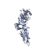
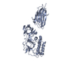
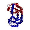
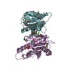
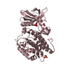
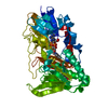
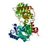
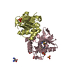
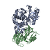
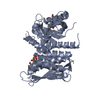
| Title | Pilus-presented adhesin, Spy0125 (Cpa), P1 form | ||||||
 Components Components | ANCILLARY PROTEIN 1 | ||||||
 Keywords Keywords | CELL ADHESION / GRAM POSITIVE PILUS / INTRAMOLECULAR ISOPEPTIDE BOND / INTERNAL THIOESTER | ||||||
| Function / homology |  Function and homology information Function and homology information: / DNA polymerase; domain 1 - #480 / Thioester domain / : / Domain of unknown function (DUF7601) / Domain of unknown function (DUF5979) / Uncharacterised domain CHP03934, TQXA / Thioester domain / Thioester domain / Prealbumin-like fold domain ...: / DNA polymerase; domain 1 - #480 / Thioester domain / : / Domain of unknown function (DUF7601) / Domain of unknown function (DUF5979) / Uncharacterised domain CHP03934, TQXA / Thioester domain / Thioester domain / Prealbumin-like fold domain / Prealbumin-like fold domain / SH3 type barrels. / DNA polymerase; domain 1 / Roll / Immunoglobulin-like fold / Immunoglobulins / Immunoglobulin-like / Sandwich / Orthogonal Bundle / Mainly Beta / Mainly Alpha Similarity search - Domain/homology | ||||||
| Biological species |  STREPTOCOCCUS PYOGENES (bacteria) STREPTOCOCCUS PYOGENES (bacteria) | ||||||
| Method |  X-RAY DIFFRACTION / SYNCHROTRON / MAD / Resolution: 1.9 Å X-RAY DIFFRACTION / SYNCHROTRON / MAD / Resolution: 1.9 Å | ||||||
 Authors Authors | Pointon, J.A. / Smith, W.D. / Saalbach, G. / Crow, A. / Kehoe, M.A. / Banfield, M.J. | ||||||
 Citation Citation | Journal: J.Biol.Chem. / Year: 2010 Title: A Highly Unusual Thioester Bond in a Pilus Adhesin is Required for Efficient Host Cell Interaction Authors: Pointon, J.A. / Smith, W.D. / Saalbach, G. / Crow, A. / Kehoe, M.A. / Banfield, M.J. | ||||||
| History |
|
- Structure visualization
| Structure viewer | Molecule: MolmilJmol/JSmol |
|---|
- Downloads & links
-Download
| PDBx/mmCIF format | 2xi9.cif.gz | 281.9 KB | Display | PDBx/mmCIF format |
|---|---|---|---|---|
| PDB format | pdb2xi9.ent.gz | 226.6 KB | Display | PDB format |
| PDBx/mmJSON format | 2xi9.json.gz | Tree view | PDBx/mmJSON format | |
| Others |  Other downloads Other downloads |
-Validation report
| Arichive directory | https://data.pdbj.org/pub/pdb/validation_reports/xi/2xi9ftp://data.pdbj.org/pub/pdb/validation_reports/xi/2xi9 | HTTPS FTP |
|---|
-Related structure data


-Links
 PDBj
PDBj- Assembly
| Deposited unit | 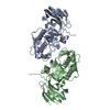
| ||||||||
|---|---|---|---|---|---|---|---|---|---|
| 1 | 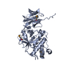
| ||||||||
| 2 | 
| ||||||||
| Unit cell |
|
-Components
| #1: Protein | Mass: 51475.434 Da / Num. of mol.: 2 / Fragment: C-TERMINAL DOMAIN, RESIDUES 286-723 Source method: isolated from a genetically manipulated source Details: INTERNAL THIOESTER LINKAGE BETWEEN CYS426 AND GLN575. INTRAMOLECULAR ISOPEPTIDE BOND BETWEEN LYS297 AND ASP595. INTRAMOLECULAR ISOPEPTIDE BOND BETWEEN LYS610 AND ASN715. Source: (gene. exp.) STREPTOCOCCUS PYOGENES (bacteria) / Strain: SF370 / Plasmid: PET28A / Production host: #2: Water | ChemComp-HOH / |  Mass: 18.015 Da / Num. of mol.: 527 / Source method: isolated from a natural source / Formula: H2O Mass: 18.015 Da / Num. of mol.: 527 / Source method: isolated from a natural source / Formula: H2OHas protein modification | Y | |
|---|
-Experimental details
-Experiment
| Experiment | Method: X-RAY DIFFRACTION / Number of used crystals: 1 |
|---|
- Sample preparation
| Crystal | Density Matthews: 2.36 Å3/Da / Density % sol: 48 % / Description: NONE |
|---|---|
| Crystal grow | pH: 5.75 Details: 38 - 40 % PEG 4000, 200 MM SODIUM ACETATE AND 100 MM TRI-SODIUM CITRATE PH 5.6 - 5.9; 34 TO 36 % PEG 5000-MME, 100 MM AMMONIUM SULPHATE, 100 MM MES PH 6.0 - 6.5 |
-Data collection
| Diffraction | Mean temperature: 100 K |
|---|---|
| Diffraction source | Source: SYNCHROTRON / Site: ESRF  / Beamline: BM14 / Wavelength: 0.9785 / Beamline: BM14 / Wavelength: 0.9785 |
| Detector | Type: ADSC CCD / Detector: CCD / Date: Jul 13, 2009 / Details: MIRROS |
| Radiation | Protocol: MAD / Monochromatic (M) / Laue (L): M / Scattering type: x-ray |
| Radiation wavelength | Wavelength: 0.9785 Å / Relative weight: 1 |
| Reflection | Resolution: 1.9→38 Å / Num. obs: 66113 / % possible obs: 93.6 % / Observed criterion σ(I): 0 / Redundancy: 7 % / Rmerge(I) obs: 0.07 / Net I/σ(I): 15.4 |
| Reflection shell | Resolution: 1.9→2 Å / Redundancy: 6.7 % / Rmerge(I) obs: 0.32 / Mean I/σ(I) obs: 5 / % possible all: 74.9 |
- Processing
| Software |
| ||||||||||||||||||||||||||||||||||||||||||||||||||||||||||||||||||||||||||||||||||||||||||||||||||||||||||||||||||||||||||||||||||||||||||||||||||||||||||||||||||||||||||||||||||||||
|---|---|---|---|---|---|---|---|---|---|---|---|---|---|---|---|---|---|---|---|---|---|---|---|---|---|---|---|---|---|---|---|---|---|---|---|---|---|---|---|---|---|---|---|---|---|---|---|---|---|---|---|---|---|---|---|---|---|---|---|---|---|---|---|---|---|---|---|---|---|---|---|---|---|---|---|---|---|---|---|---|---|---|---|---|---|---|---|---|---|---|---|---|---|---|---|---|---|---|---|---|---|---|---|---|---|---|---|---|---|---|---|---|---|---|---|---|---|---|---|---|---|---|---|---|---|---|---|---|---|---|---|---|---|---|---|---|---|---|---|---|---|---|---|---|---|---|---|---|---|---|---|---|---|---|---|---|---|---|---|---|---|---|---|---|---|---|---|---|---|---|---|---|---|---|---|---|---|---|---|---|---|---|---|
| Refinement | Method to determine structure: MAD Starting model: NONE Resolution: 1.9→34.11 Å / Cor.coef. Fo:Fc: 0.925 / Cor.coef. Fo:Fc free: 0.904 / SU B: 5.937 / SU ML: 0.08 / Cross valid method: THROUGHOUT / ESU R: 0.148 / ESU R Free: 0.135 / Stereochemistry target values: MAXIMUM LIKELIHOOD Details: HYDROGENS HAVE BEEN ADDED IN THE RIDING POSITIONS. CA 120 RESIDUES AT C_TERMINUS ARE DISORDERED. ATOM RECORD CONTAINS SUM OF TLS AND RESIDUAL B FACTORS. ANISOU RECORD CONTAINS SUM OF TLS AND ...Details: HYDROGENS HAVE BEEN ADDED IN THE RIDING POSITIONS. CA 120 RESIDUES AT C_TERMINUS ARE DISORDERED. ATOM RECORD CONTAINS SUM OF TLS AND RESIDUAL B FACTORS. ANISOU RECORD CONTAINS SUM OF TLS AND RESIDUAL U FACTORS. ATOM AND ANISO RECORDS BELOW CONTAIN THE SUM OF TLS AND RESIDUAL B/U FACTORS, AS OUPUT FROM REFMAC5 USING THE 'TLSO ADDU' OPTION. WATERS HAVE BEEN INCLUDED IN THE TLS REFINEMENT, BUT TO WHICH GROUP THEY HAVE BEEN ASSIGNED IS UNKNOWN.
| ||||||||||||||||||||||||||||||||||||||||||||||||||||||||||||||||||||||||||||||||||||||||||||||||||||||||||||||||||||||||||||||||||||||||||||||||||||||||||||||||||||||||||||||||||||||
| Solvent computation | Ion probe radii: 0.8 Å / Shrinkage radii: 0.8 Å / VDW probe radii: 1.4 Å / Solvent model: MASK | ||||||||||||||||||||||||||||||||||||||||||||||||||||||||||||||||||||||||||||||||||||||||||||||||||||||||||||||||||||||||||||||||||||||||||||||||||||||||||||||||||||||||||||||||||||||
| Displacement parameters | Biso mean: 20.65 Å2
| ||||||||||||||||||||||||||||||||||||||||||||||||||||||||||||||||||||||||||||||||||||||||||||||||||||||||||||||||||||||||||||||||||||||||||||||||||||||||||||||||||||||||||||||||||||||
| Refinement step | Cycle: LAST / Resolution: 1.9→34.11 Å
| ||||||||||||||||||||||||||||||||||||||||||||||||||||||||||||||||||||||||||||||||||||||||||||||||||||||||||||||||||||||||||||||||||||||||||||||||||||||||||||||||||||||||||||||||||||||
| Refine LS restraints |
|