 Movie
Movie Controller


+ Open data

- Basic information
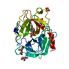
| Entry | Database: PDB / ID: 1tgn | |||||||||
|---|---|---|---|---|---|---|---|---|---|---|
| Title | STRUCTURE OF BOVINE TRYPSINOGEN AT 1.9 ANGSTROMS RESOLUTION | |||||||||
 Components Components | TRYPSINOGEN | |||||||||
 Keywords Keywords | HYDROLASE ZYMOGEN (SERINE PROTEINASE) | |||||||||
| Function / homology |  Function and homology information Function and homology informationtrypsin / serpin family protein binding / serine protease inhibitor complex / digestion / endopeptidase activity / serine-type endopeptidase activity / proteolysis / : / metal ion binding Similarity search - Function | |||||||||
| Biological species |  | |||||||||
| Method |  X-RAY DIFFRACTION / Resolution: 1.65 Å X-RAY DIFFRACTION / Resolution: 1.65 Å | |||||||||
 Authors Authors | Kossiakoff, A.A. / Stroud, R.M. | |||||||||
 Citation Citation | Journal: Biochemistry / Year: 1977 Title: Structure of bovine trypsinogen at 1.9 A resolution. Authors: Kossiakoff, A.A. / Chambers, J.L. / Kay, L.M. / Stroud, R.M. #1: Journal: Annu.Rev.Biophys.Bioeng. / Year: 1977Title: Mechanisms of Zymogen Activation Authors: Stroud, R.M. / Kossiakoff, A.A. / Chambers, J.L. | |||||||||
| History |
|
- Structure visualization
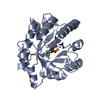
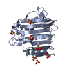
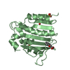
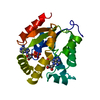
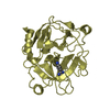
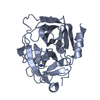
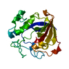
| Structure viewer | Molecule: MolmilJmol/JSmol |
|---|
- Downloads & links
-Download
| PDBx/mmCIF format | 1tgn.cif.gz | 49.6 KB | Display | PDBx/mmCIF format |
|---|---|---|---|---|
| PDB format | pdb1tgn.ent.gz | 34.5 KB | Display | PDB format |
| PDBx/mmJSON format | 1tgn.json.gz | Tree view | PDBx/mmJSON format | |
| Others |  Other downloads Other downloads |
-Validation report
| Arichive directory | https://data.pdbj.org/pub/pdb/validation_reports/tg/1tgnftp://data.pdbj.org/pub/pdb/validation_reports/tg/1tgn | HTTPS FTP |
|---|
-Related structure data
| Similar structure data |
|---|


-Links
 PDBj
PDBj

- Assembly
| Deposited unit | 
| ||||||||
|---|---|---|---|---|---|---|---|---|---|
| 1 |
| ||||||||
| Unit cell |
| ||||||||
| Atom site foot note | 1: THE AUTOLYSIS LOOP (RESIDUES 143 THROUGH 151) IS DISORDERED AND THE PRESENT INTERPRETATION IS TENTATIVE. |
-Components
| #1: Protein | Mass: 24012.953 Da / Num. of mol.: 1 Source method: isolated from a genetically manipulated source Source: (gene. exp.) |
|---|---|
| Has protein modification | Y |
-Experimental details
-Experiment
| Experiment | Method: X-RAY DIFFRACTION |
|---|
- Sample preparation
| Crystal | Density Matthews: 2 Å3/Da / Density % sol: 38.42 % | |||||||||||||||
|---|---|---|---|---|---|---|---|---|---|---|---|---|---|---|---|---|
| Crystal grow | *PLUS Temperature: 5 ℃ / Method: vapor diffusion | |||||||||||||||
| Components of the solutions | *PLUS
|
- Processing
| Refinement | Highest resolution: 1.65 Å Details: THE AUTOLYSIS LOOP (RESIDUES 143 THROUGH 151) IS DISORDERED AND THE PRESENT INTERPRETATION IS TENTATIVE. | ||||||||||||
|---|---|---|---|---|---|---|---|---|---|---|---|---|---|
| Refinement step | Cycle: LAST / Highest resolution: 1.65 Å
| ||||||||||||
| Refinement | *PLUS Rfactor obs: 0.223 | ||||||||||||
| Solvent computation | *PLUS | ||||||||||||
| Displacement parameters | *PLUS |
