 Movie
Movie Controller

[English] 日本語
 Yorodumi
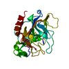
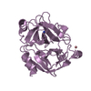
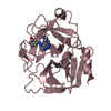
- PDB-6nvb: Crystal structure of the inhibitor-free form of the serine protea... -
+ Open data

- Basic information
| Entry | Database: PDB / ID: 6nvb | ||||||
|---|---|---|---|---|---|---|---|
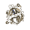
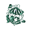
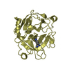
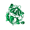
| Title | Crystal structure of the inhibitor-free form of the serine protease kallikrein-4 | ||||||
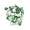
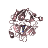
 Components Components | Kallikrein-4 | ||||||
 Keywords Keywords | HYDROLASE / protease / klk4 | ||||||
| Function / homology |  Function and homology information Function and homology informationbiomineral tissue development / amelogenesis / extracellular matrix disassembly / Hydrolases; Acting on peptide bonds (peptidases); Serine endopeptidases / secretory granule / serine-type peptidase activity / protein maturation / serine-type endopeptidase activity / proteolysis / : ...biomineral tissue development / amelogenesis / extracellular matrix disassembly / Hydrolases; Acting on peptide bonds (peptidases); Serine endopeptidases / secretory granule / serine-type peptidase activity / protein maturation / serine-type endopeptidase activity / proteolysis / : / extracellular region / metal ion binding Similarity search - Function | ||||||
| Biological species |  Homo sapiens (human) Homo sapiens (human) | ||||||
| Method |  X-RAY DIFFRACTION / SYNCHROTRON / MOLECULAR REPLACEMENT / Resolution: 1.636 Å X-RAY DIFFRACTION / SYNCHROTRON / MOLECULAR REPLACEMENT / Resolution: 1.636 Å | ||||||
 Authors Authors | Riley, B.T. / Buckle, A.M. / McGowan, S. | ||||||
 Citation Citation | Journal: Acta Crystallogr.,Sect.F / Year: 2019 Title: Crystal structure of the inhibitor-free form of the serine protease kallikrein-4. Authors: Riley, B.T. / Hoke, D.E. / McGowan, S. / Buckle, A.M. | ||||||
| History |
|
- Structure visualization
| Structure viewer | Molecule: MolmilJmol/JSmol |
|---|
- Downloads & links
-Download
| PDBx/mmCIF format | 6nvb.cif.gz | 434.9 KB | Display | PDBx/mmCIF format |
|---|---|---|---|---|
| PDB format | pdb6nvb.ent.gz | 294.7 KB | Display | PDB format |
| PDBx/mmJSON format | 6nvb.json.gz | Tree view | PDBx/mmJSON format | |
| Others |  Other downloads Other downloads |
-Validation report
| Arichive directory | https://data.pdbj.org/pub/pdb/validation_reports/nv/6nvbftp://data.pdbj.org/pub/pdb/validation_reports/nv/6nvb | HTTPS FTP |
|---|
-Related structure data
| Related structure data | 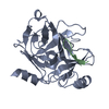 4k8yS S: Starting model for refinement |
|---|---|
| Similar structure data |

-Links
 PDBj
PDBj- Assembly
| Deposited unit | 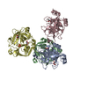
| ||||||||||||
|---|---|---|---|---|---|---|---|---|---|---|---|---|---|
| 1 | 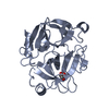
| ||||||||||||
| 2 | 
| ||||||||||||
| 3 | 
| ||||||||||||
| 4 | 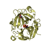
| ||||||||||||
| Unit cell |
|
-Components
| #1: Protein | Mass: 24013.090 Da / Num. of mol.: 4 Source method: isolated from a genetically manipulated source Source: (gene. exp.) Homo sapiens (human) / Gene: KLK4, EMSP1, PRSS17, PSTS / Production host:  References: UniProt: Q9Y5K2, Hydrolases; Acting on peptide bonds (peptidases); Serine endopeptidases #2: Chemical | ChemComp-GOL /   Mass: 92.094 Da / Num. of mol.: 6 / Source method: obtained synthetically / Formula: C3H8O3 Mass: 92.094 Da / Num. of mol.: 6 / Source method: obtained synthetically / Formula: C3H8O3#3: Chemical |   Mass: 96.063 Da / Num. of mol.: 2 / Source method: obtained synthetically / Formula: SO4 Mass: 96.063 Da / Num. of mol.: 2 / Source method: obtained synthetically / Formula: SO4#4: Water | ChemComp-HOH / |  Mass: 18.015 Da / Num. of mol.: 1074 / Source method: isolated from a natural source / Formula: H2O Mass: 18.015 Da / Num. of mol.: 1074 / Source method: isolated from a natural source / Formula: H2OHas protein modification | Y | |
|---|
-Experimental details
-Experiment
| Experiment | Method: X-RAY DIFFRACTION / Number of used crystals: 1 |
|---|
- Sample preparation
| Crystal | Density Matthews: 2.42 Å3/Da / Density % sol: 49.23 % |
|---|---|
| Crystal grow | Temperature: 293 K / Method: vapor diffusion, hanging drop / pH: 4.5 Details: 0.2M lithium sulfate, 0.1M sodium acetate, 22% PEG 8000 cryoprotectant = 1:1 glycerol : mother liquor |
-Data collection
| Diffraction | Mean temperature: 100 K / Ambient temp details: liquid N2 vapor stream / Serial crystal experiment: N |
|---|---|
| Diffraction source | Source: SYNCHROTRON / Site: Australian Synchrotron  / Beamline: MX1 / Wavelength: 0.9537 Å / Beamline: MX1 / Wavelength: 0.9537 Å |
| Detector | Type: ADSC QUANTUM 210r / Detector: CCD / Date: May 5, 2017 |
| Radiation | Monochromator: Si(111) / Protocol: SINGLE WAVELENGTH / Monochromatic (M) / Laue (L): M / Scattering type: x-ray |
| Radiation wavelength | Wavelength: 0.9537 Å / Relative weight: 1 |
| Reflection | Resolution: 1.636→35.8 Å / Num. obs: 107669 / % possible obs: 96.78 % / Redundancy: 3.9 % / Biso Wilson estimate: 15.75 Å2 / CC1/2: 0.999 / Rmerge(I) obs: 0.06342 / Rpim(I) all: 0.03715 / Rrim(I) all: 0.07355 / Net I/av σ(I): 17.81 / Net I/σ(I): 17.81 |
| Reflection shell | Resolution: 1.636→1.695 Å / Redundancy: 3.8 % / Rmerge(I) obs: 0.6866 / Mean I/σ(I) obs: 2.09 / Num. unique obs: 10360 / CC1/2: 0.698 / Rpim(I) all: 0.4039 / Rrim(I) all: 0.7973 / % possible all: 93.42 |
- Processing
| Software |
| |||||||||||||||||||||
|---|---|---|---|---|---|---|---|---|---|---|---|---|---|---|---|---|---|---|---|---|---|---|
| Refinement | Method to determine structure: MOLECULAR REPLACEMENT Starting model: 4K8Y Resolution: 1.636→35.8 Å / Cross valid method: FREE R-VALUE
| |||||||||||||||||||||
| Displacement parameters | Biso mean: 21.54 Å2 | |||||||||||||||||||||
| Refinement step | Cycle: LAST / Resolution: 1.636→35.8 Å
| |||||||||||||||||||||
| LS refinement shell | Resolution: 1.636→1.695 Å
|
