| 登録情報 | データベース: PDB / ID: 1lqe
|
|---|
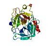
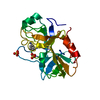
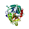
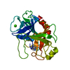
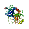
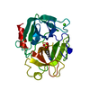
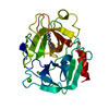
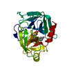
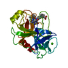
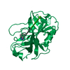
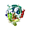
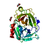
| タイトル | CRYSTAL STRUCTURE OF TRYPSIN IN COMPLEX WITH 79. |
|---|
 要素 要素 | TRYPSIN |
|---|
 キーワード キーワード | HYDROLASE / Factor Xa inhibitor / enzyme-inhibitor complex / blood coagulation factor / serine proteinase |
|---|
| 機能・相同性 |  機能・相同性情報 機能・相同性情報
trypsin / serpin family protein binding / serine protease inhibitor complex / digestion / endopeptidase activity / serine-type endopeptidase activity / proteolysis / : / metal ion binding類似検索 - 分子機能 : / Serine proteases, trypsin family, histidine active site / Serine proteases, trypsin family, serine active site / Serine proteases, trypsin family, histidine active site. / Serine proteases, trypsin family, serine active site. / Peptidase S1A, chymotrypsin family / Serine proteases, trypsin domain profile. / Trypsin-like serine protease / Serine proteases, trypsin domain / Trypsin ...: / Serine proteases, trypsin family, histidine active site / Serine proteases, trypsin family, serine active site / Serine proteases, trypsin family, histidine active site. / Serine proteases, trypsin family, serine active site. / Peptidase S1A, chymotrypsin family / Serine proteases, trypsin domain profile. / Trypsin-like serine protease / Serine proteases, trypsin domain / Trypsin / Trypsin-like serine proteases / Thrombin, subunit H / Peptidase S1, PA clan, chymotrypsin-like fold / Peptidase S1, PA clan / Beta Barrel / Mainly Beta類似検索 - ドメイン・相同性 |
|---|
| 生物種 |   Bos taurus (ウシ) Bos taurus (ウシ) |
|---|
| 手法 |  X線回折 / 分子置換 / 解像度: 2.2 Å X線回折 / 分子置換 / 解像度: 2.2 Å |
|---|
 データ登録者 データ登録者 | Schreuder, H.A. / Liesum, A. |
|---|
 引用 引用 | ジャーナル: J.Med.Chem. / 年: 2002
タイトル: Design and quantitative structure-activity relationship of 3-amidinobenzyl-1H-indole-2-carboxamides as potent, nonchiral, and selective inhibitors of blood coagulation factor Xa.
著者: Matter, H. / Defossa, E. / Heinelt, U. / Blohm, P.M. / Schneider, D. / Mueller, A. / Herok, S. / Schreuder, H.A. / Liesum, A. / Brachvogel, V. / Loenze, P. / Walser, A. / Al-Obeidi, F. / Wildgoose, P. |
|---|
| 履歴 | | 登録 | 2002年5月10日 | 登録サイト: RCSB / 処理サイト: PDBJ |
|---|
| 改定 1.0 | 2003年5月10日 | Provider: repository / タイプ: Initial release |
|---|
| 改定 1.1 | 2008年4月28日 | Group: Version format compliance |
|---|
| 改定 1.2 | 2011年7月13日 | Group: Version format compliance |
|---|
| 改定 1.3 | 2024年10月16日 | Group: Data collection / Database references ...Data collection / Database references / Derived calculations / Structure summary
カテゴリ: chem_comp_atom / chem_comp_bond ...chem_comp_atom / chem_comp_bond / database_2 / pdbx_entry_details / pdbx_modification_feature / pdbx_struct_conn_angle / struct_conn / struct_site
Item: _database_2.pdbx_DOI / _database_2.pdbx_database_accession ..._database_2.pdbx_DOI / _database_2.pdbx_database_accession / _pdbx_struct_conn_angle.ptnr1_auth_comp_id / _pdbx_struct_conn_angle.ptnr1_auth_seq_id / _pdbx_struct_conn_angle.ptnr1_label_asym_id / _pdbx_struct_conn_angle.ptnr1_label_atom_id / _pdbx_struct_conn_angle.ptnr1_label_comp_id / _pdbx_struct_conn_angle.ptnr1_label_seq_id / _pdbx_struct_conn_angle.ptnr3_auth_comp_id / _pdbx_struct_conn_angle.ptnr3_auth_seq_id / _pdbx_struct_conn_angle.ptnr3_label_asym_id / _pdbx_struct_conn_angle.ptnr3_label_atom_id / _pdbx_struct_conn_angle.ptnr3_label_comp_id / _pdbx_struct_conn_angle.ptnr3_label_seq_id / _pdbx_struct_conn_angle.value / _struct_conn.pdbx_dist_value / _struct_conn.ptnr1_auth_comp_id / _struct_conn.ptnr1_auth_seq_id / _struct_conn.ptnr1_label_asym_id / _struct_conn.ptnr1_label_atom_id / _struct_conn.ptnr1_label_comp_id / _struct_conn.ptnr1_label_seq_id / _struct_conn.ptnr2_auth_comp_id / _struct_conn.ptnr2_auth_seq_id / _struct_conn.ptnr2_label_asym_id / _struct_conn.ptnr2_label_atom_id / _struct_conn.ptnr2_label_comp_id / _struct_conn.ptnr2_label_seq_id / _struct_site.pdbx_auth_asym_id / _struct_site.pdbx_auth_comp_id / _struct_site.pdbx_auth_seq_id |
|---|
|
|---|
 ムービー
ムービー コントローラー


 データを開く
 基本情報
基本情報 構造の表示
構造の表示 ダウンロードとリンク
 その他のダウンロード


 PDBj
PDBj

 集合体
集合体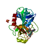

 分子量: 40.078 Da / 分子数: 1 / 由来タイプ: 合成 / 式: Ca
 分子量: 96.063 Da / 分子数: 1 / 由来タイプ: 合成 / 式: SO4
 分子量: 546.682 Da / 分子数: 1 / 由来タイプ: 合成 / 式: C34H36N5O2
 分子量: 18.015 Da / 分子数: 139 / 由来タイプ: 天然 / 式: H2O
 試料調製
試料調製 解析
解析