 Movie
Movie Controller

[English] 日本語
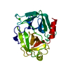
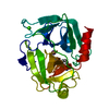
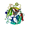
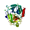
 Yorodumi
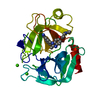
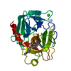
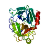
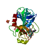
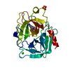
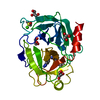
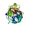
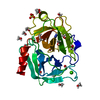
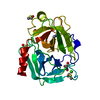
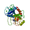
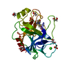
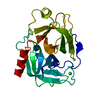
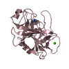
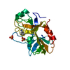
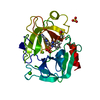
- PDB-1qb1: Bovine Trypsin with 1-[2-[5-[amino(imino)methyl]-2-hydroxyphenoxy... -
+ Open data

- Basic information
| Entry | Database: PDB / ID: 1qb1 | ||||||
|---|---|---|---|---|---|---|---|
| Title | Bovine Trypsin with 1-[2-[5-[amino(imino)methyl]-2-hydroxyphenoxy]-6-[3-(4,5-dihydro-1-methyl-1H-imidazol-2-yl)phenoxy]pyridin-4-yl]piperidine-3-carboxylic Acid (ZK-806974) | ||||||
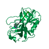
 Components Components | PROTEIN (TRYPSIN) | ||||||
 Keywords Keywords | HYDROLASE / SERINE PROTEINASE / PROTEIN-INHIBITOR COMPLEX / S1 POCKET / FACTOR XA | ||||||
| Function / homology |  Function and homology information Function and homology informationtrypsin / serpin family protein binding / serine protease inhibitor complex / digestion / endopeptidase activity / serine-type endopeptidase activity / proteolysis / : / metal ion binding Similarity search - Function | ||||||
| Biological species |  | ||||||
| Method |  X-RAY DIFFRACTION / DIRECT REPLACEMENT / Resolution: 1.8 Å X-RAY DIFFRACTION / DIRECT REPLACEMENT / Resolution: 1.8 Å | ||||||
 Authors Authors | Whitlow, M. | ||||||
 Citation Citation | Journal: Acta Crystallogr.,Sect.D / Year: 1999 Title: Crystallographic analysis of potent and selective factor Xa inhibitors complexed to bovine trypsin. Authors: Whitlow, M. / Arnaiz, D.O. / Buckman, B.O. / Davey, D.D. / Griedel, B. / Guilford, W.J. / Koovakkat, S.K. / Liang, A. / Mohan, R. / Phillips, G.B. / Seto, M. / Shaw, K.J. / Xu, W. / Zhao, Z. ...Authors: Whitlow, M. / Arnaiz, D.O. / Buckman, B.O. / Davey, D.D. / Griedel, B. / Guilford, W.J. / Koovakkat, S.K. / Liang, A. / Mohan, R. / Phillips, G.B. / Seto, M. / Shaw, K.J. / Xu, W. / Zhao, Z. / Light, D.R. / Morrissey, M.M. #1: Journal: FEBS Lett. / Year: 1978Title: Crystal Structure Analysis and Refinement of Two Variants of Trigonal Trypsinogen Authors: Bode, W. / Huber, R. #2: Journal: J.Med.Chem. / Year: 1999Title: Design, Synthesis, and Activity of 2,6-Diphenoxypyridine-Derived Factor Xa Inhibitors Authors: Phillips, G. / Davey, D. / Eagen, K. / Ng, H.P. / Pinkerton, M. / Koovakkat, S.K. / Whitlow, M. / Liang, A. / Trinh, L. / Morrissey, M.M. #3: Journal: J.Med.Chem. / Year: 1998Title: Discovery of N-[2-[5-[Amino(Imino)Methyl]-2-Hydroxyphenoxy]-3,5- Difluoro-6-[3-(4,5-Dihydro-1-Methyl-1H-Imidazol-2-Yl)Phenoxy] Pyridin-4-Yl-N-Methylglycine (Zk-807834): A Potent, Selective and ...Title: Discovery of N-[2-[5-[Amino(Imino)Methyl]-2-Hydroxyphenoxy]-3,5- Difluoro-6-[3-(4,5-Dihydro-1-Methyl-1H-Imidazol-2-Yl)Phenoxy] Pyridin-4-Yl-N-Methylglycine (Zk-807834): A Potent, Selective and Orally Active Inhibitor of the Blood Coagulation Enzyme Factor Xa Authors: Phillips, G.B. / Buckman, B.O. / Davey, D.D. / Eagen, K.A. / Guilford, W.J. / Hinchman, J. / Ho, E. / Koovakkat, S. / Liang, A. / Light, D.R. / Mohan, R. / Ng, H.P. / Post, J. / Smith, D. / ...Authors: Phillips, G.B. / Buckman, B.O. / Davey, D.D. / Eagen, K.A. / Guilford, W.J. / Hinchman, J. / Ho, E. / Koovakkat, S. / Liang, A. / Light, D.R. / Mohan, R. / Ng, H.P. / Post, J. / Smith, D. / Subramanyam, B. / Sullivan, M.E. / Trinh, L. / Vergona, R. / Walters, J. / White, K. / Whitlow, M. / Wu, S. / Xu, W. / Morrissey, M.M. #4: Journal: FEBS Lett. / Year: 1995Title: Crystal Structures of Factor Xa Specific Inhibitors in Complex with Trypsin: Structural Grounds for Inhibition of Factor Xa and Selectivity Against Thrombin Authors: Stubbs, M.T. / Huber, R. / Bode, W. #5: Journal: J.Biol.Chem. / Year: 1996Title: X-Ray Structure of Active Site-Inhibited Clotting Factor Xa. Implications for Drug Design and Substrate Recognition Authors: Brandstetter, H. / Kuhne, A. / Bode, W. / Huber, R. / Von Der Saal, W. / Wirthensohn, K. / Engh, R.A. #6: Journal: J.Mol.Biol. / Year: 1993Title: Structure of Human Des(1-45) Factor Xa at 2.2 A Resolution Authors: Padmanabhan, K. / Padmanabhan, K.P. / Tulinsky, A. / Park, C.H. / Bode, W. / Huber, R. / Blankenship, D.T. / Cardin, A.D. / Kisiel, W. #7: Journal: Eur.J.Biochem. / Year: 1990Title: Geometry of Binding of the Benzamidine-and Arginine-Based Inhibitors N Alpha-(2-Naphthyl-Sulphonyl-Glycyl)-Dl-P-Amidinophenylalanyl-Piperidine (Napap) and (2R,4R)-4-Methyl-1-[N Alpha-(3-Methyl- ...Title: Geometry of Binding of the Benzamidine-and Arginine-Based Inhibitors N Alpha-(2-Naphthyl-Sulphonyl-Glycyl)-Dl-P-Amidinophenylalanyl-Piperidine (Napap) and (2R,4R)-4-Methyl-1-[N Alpha-(3-Methyl-1,2,3,4-Tetrahydro-8-Quinolinesulphonyl)- L-Arginyl]-2-Piperidine to Carboxylic Acid (Mqpa) Human Alpha-Thrombin. X-Ray Crystallographic Determination of the Napap-Trypsin Complex and Modeling of Napap-Thrombin and Mqpa-Thrombin Authors: Bode, W. / Turk, D. / Sturzebecher, J. #8: Journal: J.Mol.Biol. / Year: 1989Title: Crystal Structure of Bovine Beta-Trypsin at 1.5 A Resolution in a Crystal Form with Low Molecular Packing Density. Active Site Geometry, Ion Pairs and Solvent Structure Authors: Bartunik, H.D. / Summers, L.J. / Bartsch, H.H. | ||||||
| History |
|
- Structure visualization
| Structure viewer | Molecule: MolmilJmol/JSmol |
|---|
- Downloads & links
-Download
| PDBx/mmCIF format | 1qb1.cif.gz | 63.1 KB | Display | PDBx/mmCIF format |
|---|---|---|---|---|
| PDB format | pdb1qb1.ent.gz | 45.2 KB | Display | PDB format |
| PDBx/mmJSON format | 1qb1.json.gz | Tree view | PDBx/mmJSON format | |
| Others |  Other downloads Other downloads |
-Validation report
| Arichive directory | https://data.pdbj.org/pub/pdb/validation_reports/qb/1qb1ftp://data.pdbj.org/pub/pdb/validation_reports/qb/1qb1 | HTTPS FTP |
|---|
-Related structure data


-Links
 PDBj
PDBj

- Assembly
| Deposited unit | 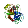
| ||||||||||
|---|---|---|---|---|---|---|---|---|---|---|---|
| 1 |
| ||||||||||
| Unit cell |
| ||||||||||
| Details | The biological assembly is a monomer. |
-Components
| #1: Protein | Mass: 23324.287 Da / Num. of mol.: 1 / Fragment: BOVINE TRYPSIN / Source method: isolated from a natural source / Details: BOEHRINGER MANNHEIM CATALOG NUMBER 109,827 / Source: (natural) |
|---|---|
| #2: Chemical | ChemComp-CA /   Mass: 40.078 Da / Num. of mol.: 1 / Source method: obtained synthetically / Formula: Ca Mass: 40.078 Da / Num. of mol.: 1 / Source method: obtained synthetically / Formula: Ca |
| #3: Chemical | ChemComp-974 /   Mass: 565.548 Da / Num. of mol.: 1 / Source method: obtained synthetically / Formula: C28H27F2N6O5 Mass: 565.548 Da / Num. of mol.: 1 / Source method: obtained synthetically / Formula: C28H27F2N6O5 |
| #4: Water | ChemComp-HOH /  Mass: 18.015 Da / Num. of mol.: 218 / Source method: isolated from a natural source / Formula: H2O Mass: 18.015 Da / Num. of mol.: 218 / Source method: isolated from a natural source / Formula: H2O |
| Has protein modification | Y |
-Experimental details
-Experiment
| Experiment | Method: X-RAY DIFFRACTION / Number of used crystals: 1 |
|---|
- Sample preparation
| Crystal | Density Matthews: 3 Å3/Da / Density % sol: 58.4 % Description: STARTING FROM THE TRYPSIN 2-AMINOBENZIMIDAZOLE STRUCTURE | ||||||||||||||||||||||||||||||||||||
|---|---|---|---|---|---|---|---|---|---|---|---|---|---|---|---|---|---|---|---|---|---|---|---|---|---|---|---|---|---|---|---|---|---|---|---|---|---|
| Crystal grow | Temperature: 297 K / Method: vapor diffusion, hanging drop / pH: 7.4 Details: MICROCRYSTALS WERE INITIALLY GROWN USING THE HANGING DROP METHOD IN LINBRO CULTURE PLATES. REFERENCES 7 (BODE, TURK & STURZEBACHER) AND 8 (BARTUNIK, SUMMERS & BARTSH) HAVE DESCRIBED THE ...Details: MICROCRYSTALS WERE INITIALLY GROWN USING THE HANGING DROP METHOD IN LINBRO CULTURE PLATES. REFERENCES 7 (BODE, TURK & STURZEBACHER) AND 8 (BARTUNIK, SUMMERS & BARTSH) HAVE DESCRIBED THE CRYSTALLIZATION OF THE ORTHORHOMBIC FORM OF BOVINE TRYPSIN. CRYSTALS WERE SEEDING INTO DROPS CONTAINING 40 MG/ML BOVINE TRYPSIN, 0.0125 M BENZAMIDINE, 0.7 TO 0.9 M AS, 0.05 M MES PH 6.0, 0.0025 M CACL2 AFTER EQUILIBRATION AGAINST THE CRYSTALLIZATION RESERVOIR FOR 1 DAY. THE CRYSTALLIZATION RESERVOIRS CONTAINED 1.4 TO 1.8 M AS, 0.1 M MES PH 6.0, 0.005 M CACL2., pH 7.4, VAPOR DIFFUSION, HANGING DROP, temperature 297K | ||||||||||||||||||||||||||||||||||||
| Crystal | *PLUS | ||||||||||||||||||||||||||||||||||||
| Crystal grow | *PLUS | ||||||||||||||||||||||||||||||||||||
| Components of the solutions | *PLUS
|
-Data collection
| Diffraction | Mean temperature: 297 K |
|---|---|
| Diffraction source | Source: ROTATING ANODE / Type: SIEMENS / Wavelength: 1.5418 |
| Detector | Type: MARRESEARCH / Detector: IMAGE PLATE / Date: Apr 12, 1995 / Details: MIRRORS |
| Radiation | Protocol: SINGLE WAVELENGTH / Monochromatic (M) / Laue (L): M / Scattering type: x-ray |
| Radiation wavelength | Wavelength: 1.5418 Å / Relative weight: 1 |
| Reflection | Resolution: 1.8→10 Å / Num. obs: 26263 / % possible obs: 99 % / Observed criterion σ(F): 0 / Observed criterion σ(I): 2 / Redundancy: 3.4 % / Rsym value: 0.078 / Net I/σ(I): 16.2 |
| Reflection shell | Resolution: 1.8→1.9 Å / Redundancy: 3.11 % / Mean I/σ(I) obs: 3.11 / Rsym value: 0.222 / % possible all: 99.9 |
| Reflection | *PLUS Rmerge(I) obs: 0.078 |
| Reflection shell | *PLUS Highest resolution: 1.8 Å / Lowest resolution: 1.9 Å / % possible obs: 99.9 % / Rmerge(I) obs: 0.222 |
- Processing
| Software |
| ||||||||||||||||||||||||||||||||||||||||||||||||||||||||||||||||||||||||||||||||||||
|---|---|---|---|---|---|---|---|---|---|---|---|---|---|---|---|---|---|---|---|---|---|---|---|---|---|---|---|---|---|---|---|---|---|---|---|---|---|---|---|---|---|---|---|---|---|---|---|---|---|---|---|---|---|---|---|---|---|---|---|---|---|---|---|---|---|---|---|---|---|---|---|---|---|---|---|---|---|---|---|---|---|---|---|---|---|
| Refinement | Method to determine structure: DIRECT REPLACEMENT / Resolution: 1.8→10 Å / Cross valid method: THROUGHOUT / σ(F): 2 Stereochemistry target values: W.A.Hendrickson (1985) Methods in Enzymology, Volume 115, pages 370-381.
| ||||||||||||||||||||||||||||||||||||||||||||||||||||||||||||||||||||||||||||||||||||
| Refinement step | Cycle: LAST / Resolution: 1.8→10 Å
| ||||||||||||||||||||||||||||||||||||||||||||||||||||||||||||||||||||||||||||||||||||
| Refine LS restraints |
| ||||||||||||||||||||||||||||||||||||||||||||||||||||||||||||||||||||||||||||||||||||
| Software | *PLUS Name: PROFFT / Classification: refinement | ||||||||||||||||||||||||||||||||||||||||||||||||||||||||||||||||||||||||||||||||||||
| Refinement | *PLUS σ(F): 2 / % reflection Rfree: 4 % / Rfactor obs: 0.183 / Rfactor Rwork: 0.183 | ||||||||||||||||||||||||||||||||||||||||||||||||||||||||||||||||||||||||||||||||||||
| Solvent computation | *PLUS | ||||||||||||||||||||||||||||||||||||||||||||||||||||||||||||||||||||||||||||||||||||
| Displacement parameters | *PLUS | ||||||||||||||||||||||||||||||||||||||||||||||||||||||||||||||||||||||||||||||||||||
| Refine LS restraints | *PLUS
|
